
About Article
Research article • Volume 1, Issue 1, Pages 10–15 (2026) 100002
Computational and Biophysical Characterization of Limonene as a Potential Natural Inhibitor of CDK6 for Therapeutic Targeting of Cancer
https://doi.org/10.53295/cmb.v1.i1.100002
Under a Creative Commons license
Open access
Abstract
Cyclin-dependent kinase 6 (CDK6) plays a central role in G1–S phase cell cycle progression and is frequently dysregulated in various cancers, making it an established therapeutic target. Although selective CDK4/6 inhibitors are clinically available, exploration of natural compounds targeting CDK6 remains limited. The present study aimed to investigate the binding mechanism and inhibitory potential of limonene against CDK6 using integrated computational and experimental approaches. Recombinant CDK6 was cloned, expressed, and purified, followed by molecular docking, fluorescence spectroscopy, and kinase inhibition assay. Docking analysis revealed a binding free energy of with a calculated of 4.62 and ligand efficiency of per non-hydrogen atom. Fluorescence quenching studies demonstrated strong binding affinity (), while enzymatic assays confirmed dose-dependent suppression of CDK6 activity. Collectively, these findings indicate that limonene directly interacts with and functionally inhibits CDK6, highlighting its potential as a natural scaffold for the development of CDK6-targeted anticancer therapeutics.
Keywords: Cyclin-dependent kinase 6, Limonene, Molecular docking, Fluorescence spectroscopy, Kinase inhibition assay, Protein-ligand interaction, Cell cycle regulation, Anticancer therapy
Introduction
Cancer is characterized by uncontrolled cell proliferation, primarily driven by the dysregulation of the cell cycle. The cell cycle comprises a series of phases, and key regulators include cyclins, cyclin-dependent kinases (CDKs), and CDK inhibitors (CKIs), which work together to control progression through these phases. The disruption of these components of the cell cycle is a core hallmark of cancer, enabling uncontrolled cell proliferation and tumorigenesis (Hanahan and Weinberg, 2011).
Cyclin-dependent kinases (CDKs) are serine/threonine protein kinases that are involved in the cell cycle, transcription, and other biological processes like translation, neurogenesis, and apoptosis (Malumbres, 2014). Among CDK family members, cyclin-dependent kinase 6 (CDK6) and CDK4 play important roles in mammalian cell proliferation by driving cells into the DNA synthetic (S) phase of the cell division cycle (Sherr et al., 2016). CDK4 and CDK6 are associated with D-type cyclins (D1, D2, and D3), which phosphorylate retinoblastoma (Rb), thereby releasing E2F transcription factors. E2F-dependent gene activation promotes G1 to S phase progression and DNA synthesis facilitating cell cycle progression (Classon and Harlow, 2002) through the early G1 phase of the cell cycle (Nebenfuehr et al., 2020). Beyond its function in cell cycle control, CDK6 contributes to cell differentiation, tissue development, and hematopoietic lineage commitment (Tigan et al., 2016). Emerging evidence also suggests that CDK6 modulates anti-tumour immunity by regulating immune cell proliferation and immune checkpoint molecule expression (Petroni et al., 2020). Figure illustrates the transcriptional, post-transcriptional, and post-translational regulation of Cyclin D–CDK4/6.
CDK6 and CDK4 form larger protein complexes with heat shock protein 90 (Hsp90) and its co-chaperone cell division cycle 37 (Cdc37), which facilitate correct protein folding, assembly, maturation, and stability of many proteins (Hallett et al., 2017). Hyperactivation of CDK6 has been linked to various malignancies, including breast cancer, leukemia, lymphoma, and glioblastoma. Experimental disruption of CDK6 prolongs the G1 phase and inhibits S-phase entry. G1-phase prolongation causes premature differentiation in neuroepithelial cells, which explains reduced proliferation after CDK6 loss (O'Sullivan, 2026).
Structurally, CDK6 is a 326-amino-acid protein (36.5 kDa) encoded on chromosome 7 (Russo et al., 1998). It comprises several key domains: the N-terminal lobe (residues 1–98) contains a regulatory cyclin-binding domain, while the C-terminal lobe forms the catalytic core (Sielecki et al., 2000). The activation T-loop (residues 156–172) within the catalytic domain regulates kinase activity through phosphorylation, while additional regulatory motifs, including a C-terminal PEST sequence, contribute to protein stability (Li et al., 2015). CDK6 activation requires interaction with cyclin D and phosphorylation by CDK-activating kinase (CAK), whereas its activity is negatively controlled by INK4 and Cip/Kip family inhibitors. Beyond phosphorylation, CDK6 is finely regulated at transcriptional, post-transcriptional (e.g., microRNAs such as miR-124 and miR-34a), and post-translational levels, including phosphorylation, acetylation, and ubiquitination (Bachs et al., 2018).
Due to its primary function in tumour growth and survival, CDK6 has been recognized as a validated therapeutic target (Yousuf et al., 2022). Selective CDK4/6 inhibitors, palbociclib, ribociclib, and abemaciclib, have shown considerable clinical benefit and have been approved by regulators for hormone receptor-positive, HER2-negative metastatic breast cancer (Rencuzogullari et al., 2020). These substances competitively bind the ATP-binding pocket of CDK4/6, inhibit Rb phosphorylation, and induce G1 cell cycle arrest. Combination strategies integrating CDK6 inhibitors with endocrine therapy, chemotherapy, or immunotherapy have further enhanced therapeutic efficacy and addressed resistance mechanisms. CDK6 disruption prolonged the G1 phase and inhibited S-phase entry in these cells. G1-phase lengthening has been shown to induce premature differentiation in neuroepithelial cells, explaining the decreased proliferation associated with CDK6 disruption (Shigenaka et al., 2025).
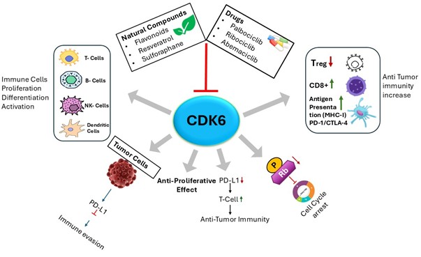
Natural products have also gained interest as potential modulators of CDK6 activity (Figure). Bioactive phytochemicals that modulate cell cycle and apoptosis-related pathways, such as flavonoids (e.g., quercetin, fisetin), resveratrol, curcumin, sulforaphane, and monoterpenes like limonene, exhibit anti-proliferative properties (Srivastava and Saxena, 2025). These compounds affect cancer-associated signaling networks, including Rb–E2F, PI3K/Akt/mTOR, NF-, and MAPK pathways, and may contribute to CDK6 inhibition either directly or indirectly (Kim et al., 2013).
Limonene, a naturally occurring monocyclic monoterpene abundant in citrus oils, promotes the GST (Glutathione S-transferase) system in the liver and small bowel, which helps in eliminating carcinogens (Sun, 2007). Limonene 1,2-diol has been reported to inhibit tumor growth through inhibition of p21-dependent signaling, induce apoptosis via induction of the transforming growth factor beta-signaling pathway, inhibit post-translational modification of signal transduction proteins, cause G1 cell cycle arrest, and modulate genes associated with cell cycle progression and apoptosis (Sun, 2007).
Collectively, CDK6 occupies a central position at the intersection of cell cycle control, oncogenic signaling, and immune regulation. Its structural features, multilayered regulatory mechanisms, and proven druggability highlight its significance as both a mechanistic driver of tumorigenesis and a compelling therapeutic target. Continued exploration of CDK6 biology and inhibition strategies, synthetic and natural, holds substantial promise for advancing precision oncology.
Figure 1. The schematic illustration of transcriptional, post-transcriptional, and post-translational regulation of Cyclin D-CDK4/6. In normal cells, controlled CDK4/6 activation leads to the regulated phosphorylation of the retinoblastoma protein (Rb), releasing E2F to permit orderly G1-S progression. In contrast, hyperactivation of CDK4/6 in cancer cells leads to excessive Rb phosphorylation, sustained E2F hyperactivation, and uncontrolled cell cycle progression, contributing to tumor development.
Download: Download full-size image
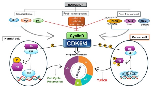
Figure 2. The diagram illustrates the central role of CDK6 in coordinating tumor cell proliferation and immune regulation. CDK6 promotes cell cycle progression by phosphorylating Rb, facilitating tumor growth and immune evasion via PD-L1 regulation. Inhibition of CDK6 by approved inhibitors or bioactive natural compounds induces cell-cycle arrest, reduces PD-L1 expression, and suppresses tumour growth. In parallel, CDK6 modulation influences immune cell proliferation, differentiation, and activation, including those of T cells, B cells, NK cells, and dendritic cells. Therapeutic targeting of CDK6 decreases regulatory T cells (Tregs) while enhancing CD8 T-cell responses and antigen presentation (MHC-I), thereby strengthening anti-tumour immunity.
Download: Download full-size image
2. Materials and Methods
2.1. Chemicals and reagents
The expression vector pET-28a(+) and Escherichia coli BL21 (DE3) competent cells were procured from Qiagen, while E. coli DH5 cells were obtained from Invitrogen. Analytical-grade reagents, including sodium chloride (NaCl), ethylenediaminetetraacetic acid (EDTA), and other routine chemicals, were purchased from Merck (India). Luria–Bertani (LB) broth was obtained from Merck (Darmstadt, Germany). Kanamycin and isopropyl--D-thiogalactopyranoside (IPTG) were sourced from Sigma-Aldrich (St. Louis, MO, USA). Nickel–nitrilotriacetic acid (Ni–NTA) affinity chromatography columns and Ni–NTA agarose beads were acquired from Bio-Rad and Qiagen (QIAexpress), respectively. Limonene used in this study was purchased from Sigma-Aldrich (USA).
2.2. Molecular docking
Molecular docking was performed to investigate the binding interactions between limonene and the crystal structure of CDK6 (PDB ID: 3NUP). Docking simulations were conducted using InstaDock, an automated platform for structure-based virtual screening (Mohammad et al., 2021). Binding affinity calculations were carried out using QuickVina-W (Hassan et al., 2017), a modified version of AutoDock Vina (Trott and Olson, 2010) that integrates empirical and knowledge-based scoring functions. A blind docking approach was employed to allow unbiased exploration of potential ligand-binding sites across the protein surface. The predicted binding free energy () obtained from docking was used to calculate the inhibitory constant () and values according to the following thermodynamic relationships:
where represents the docking-derived binding free energy, R is the universal gas constant (), T is the temperature (298.15 K), and is the predicted inhibition constant (Shityakov, 2014).
To further evaluate ligand binding efficiency, ligand efficiency (LE) was calculated using:
where LE denotes ligand efficiency ( per non-hydrogen atom), is the binding free energy, and N corresponds to the number of non-hydrogen atoms in the ligand. This parameter provides a normalized assessment of binding strength relative to molecular size (Hopkins et al., 2004).
2.3. Plasmid isolation
The overnight bacterial culture was pelleted by centrifugation at 6000 rpm for 5 minutes at room temperature, then resuspended in the resuspension buffer. Lysis was performed to release cellular contents, including plasmid DNA, using the lysis buffer. Proteins were then digested using proteinase K. Purification of the plasmid was done using a phenol–chloroform extraction kit. Finally, the concentration and purity of plasmid DNA were checked using a NanoDrop at 260 nm. Purified bands of isolated plasmid were also confirmed using agarose gel electrophoresis.
2.4. Agarose gel electrophoresis
A 0.8% agarose gel was prepared by adding TAE buffer and heating in a microwave. EtBr was added to the cooled gel solution, and then the cooled gel was poured into the gel tray. Gel was allowed to solidify for about 20 min. Samples were prepared by adding loading dye (bromophenol blue) to the isolated plasmid DNA, and loading the samples into the wells after removing the comb. The tank was filled with TAE buffer, and the leads were connected to the power supply. After applying a constant voltage of 100–150 V for about 45–60 minutes, the gel was removed from the gel box, placed on a UV transilluminator, and visualized using a gel documentation system.
2.5. Competent cell preparation and transformation
A single colony was inoculated into LB broth containing kanamycin and incubated overnight at C, then transferred to a larger volume of fresh LB broth and incubated until the OD reached 0.4–0.6 at 600 nm. Metabolic activity was reduced by cooling the culture on ice for 15–30 minutes. The culture was centrifuged, and the supernatant was discarded. The pellet was resuspended in ice-cold 10% glycerol solution. The competent cells formed were aliquoted into microcentrifuge tubes and stored at C (Bird et al., 2022).
The recombinant plasmid construct pET-28a(+) harboring the CDK6 gene was transformed into Escherichia coli BL21 (DE3) competent cells using the heat-shock method. Briefly, chemically competent BL21 (DE3) cells were thawed on ice, and 50–100 ng of plasmid DNA was gently mixed with the cells. The mixture was incubated on ice for 20 min to facilitate DNA adsorption. Subsequently, cells were subjected to a heat shock at C for 90 s, followed by immediate ice incubation for 5 min to stabilize the membranes. Thereafter, of Luria–Bertani (LB) broth was added, and the cells were allowed to recover at C with shaking at 220 rpm for 1 h. The transformed cells were then spread onto LB agar plates supplemented with kanamycin and incubated overnight (12–16 h) at C to allow colony formation (Yousuf et al., 2022).
2.6. Expression and purification
The full-length CDK6 gene "981-bp" coding sequence was cloned into the pET-28a(+) expression vector and sequence-verified prior to protein expression. The recombinant plasmid was transformed into Escherichia coli BL21 (DE3) cells for heterologous protein production. Transformed cells were cultured in LB medium containing kanamycin at C until the desired optical density was reached, then induced with 0.25 mM isopropyl--D-thiogalactopyranoside (IPTG). After induction, cells were harvested by centrifugation and resuspended in lysis buffer (25 mM Tris–HCl, pH 8.0, 200 mM NaCl, 1 mM DTT, and 10 mM PMSF). Cell disruption was performed by sonication on ice, and the lysate was centrifuged at 9,000 rpm for 20 min at C to separate soluble and insoluble fractions. The pellet containing inclusion bodies was collected and washed three times with Milli-Q water to remove impurities.
The inclusion bodies were subsequently solubilized using a buffer containing 25 mM Tris–HCl, 200 mM NaCl, and 0.5% sarcosine and loaded onto a pre-equilibrated Ni–NTA affinity chromatography column for purification. After binding, the column was washed to remove non-specifically bound proteins, and recombinant CDK6 was eluted using elution buffer containing 150 mM imidazole (25 mM Tris–HCl, pH 8.0, 200 mM NaCl). Protein purity and molecular weight were analyzed using 12% SDS–PAGE (Yousuf et al., 2020).
2.7. Kinase inhibition assay
The inhibitory effect of limonene on recombinant CDK6 was evaluated using a malachite green–based ATPase assay. The reaction mixture (final volume: ) contained purified CDK6 () and freshly prepared ATP (). Increasing concentrations of limonene were added to assess dose-dependent inhibition. The reaction mixtures were incubated at C for 45 min to allow ATP hydrolysis. The reaction was terminated by adding of malachite green reagent, followed by incubation at room temperature for 15–20 min to enable color development resulting from inorganic phosphate release. Absorbance was measured at 600 nm using a microplate ELISA reader in a 96-well format. Enzyme activity was calculated based on phosphate release, and percentage inhibition was determined relative to control reactions lacking inhibitor (Voura et al., 2019).
2.8. Fluorescence measurement
The interaction between limonene and purified recombinant CDK6 was investigated using intrinsic fluorescence spectroscopy by monitoring changes in the emission spectrum of CDK6 upon ligand titration (Jameel et al., 2017). Fluorescence measurements were performed using a JASCO spectrofluorometer (Model FP-6200). Titration experiments were conducted by progressively increasing the limonene concentration, and each measurement was performed in triplicate to ensure reproducibility. Changes in fluorescence intensity were recorded and analyzed to determine binding parameters. The binding constant (), number of binding sites (n), and associated thermodynamic parameters were calculated from fluorescence quenching data using the Stern–Volmer equation. Analysis of the quenching behavior enabled characterization of the binding affinity and interaction mechanism between limonene and CDK6 (Shamsi et al., 2020).
3. Results
3.1. Molecular docking
Docking analysis of Conformer3D_COMPOUND_CID_22311 (limonene) against the CDK6 crystal structure (PDB ID: 3NUP) revealed a predicted binding free energy () of . The corresponding calculated value was 4.62, indicating moderate binding affinity toward the target protein. The ligand efficiency (LE) was determined to be per non-hydrogen atom, suggesting favorable binding energy relative to molecular size (Figure 3).
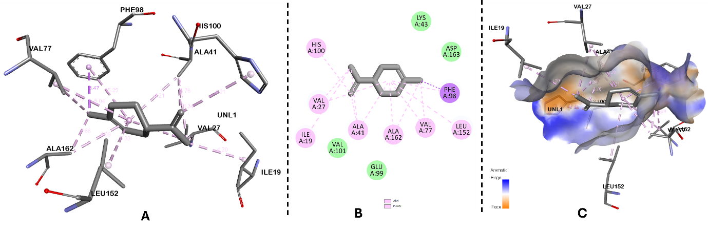
Comprehensive interaction profiling of all generated docked conformations was performed to examine the binding orientation and molecular interactions within the 3NUP binding pocket (Figure 4). Analysis of the best-ranked pose revealed that the ligand occupies the active-site cavity and establishes stabilizing interactions with key amino acid residues, thereby contributing to its predicted binding affinity.
Figure 3. Representation of the binding mode of limonene to the CDK6. (A) Cartoon representation of CDK6 in complex with docked limonene. (B) Zoomed interaction of limonene with the active-site pocket residues of CDK6.
Download: Download full-size image
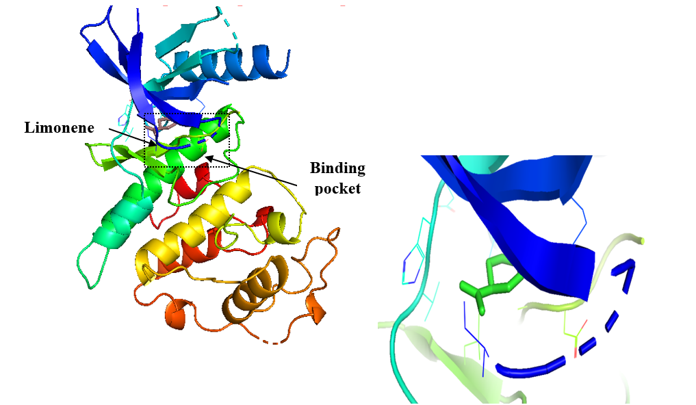
Figure 4. Molecular docking analysis of limonene with CDK6 active site. (A) Three-dimensional binding pose of limonene in the CDK6 catalytic pocket, showing key residues involved in hydrophobic stabilization. (B) Two-dimensional map showing hydrogen bonding and -alkyl/alkyl interactions between limonene and active site amino acids. (C) Surface representation of the CDK6 binding pocket illustrating favourable shape complementarity and hydrophobic interactions.
Download: Download full-size image
3.2. Protein expression and purification
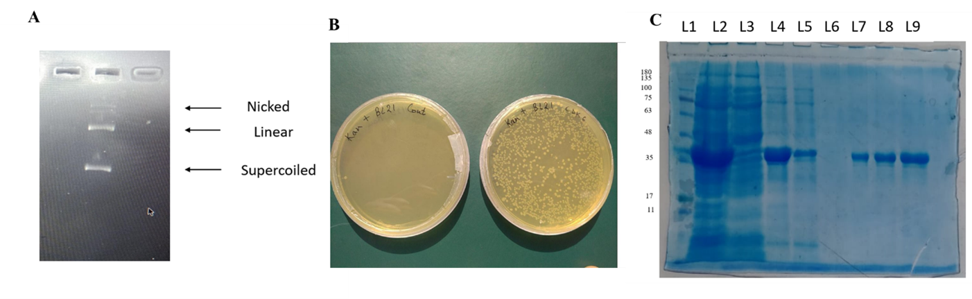
Plasmid isolated using the QIAprep Spin Miniprep Kit was checked for purity and concentration using a Nanodrop, which showed a purity of about 1.8 (A_260/280), and digested bands were visualized by running an agarose gel, which showed clear bands for nicked, linear, and supercoiled forms of DNA (Figure 5A). The confirmed pET28a+ plasmid was successfully transformed into E. coli strain BL21 (DE3). Colonies appeared after incubation with kanamycin at 50 µg/mL for 12–16 hours at C (Figure 5B). Recombinant CDK6 protein was expressed at C by inducing with 0.25 mM IPTG. Purified CDK6 protein was obtained by Ni–NTA affinity chromatography, confirmed by running SDS–PAGE (Figure 5C).
Figure 5. (A) Restriction digestion of the isolated plasmid, lane 1 showing the nicked, linear, and supercoiled form of DNA. (B) Transformed E. coli BL21 colonies on an LB agar plate. (C) Purified CDK6 protein, eluted with 150 mM imidazole-containing elution buffer.
Download: Download full-size image
3.3. Fluorescence binding studies
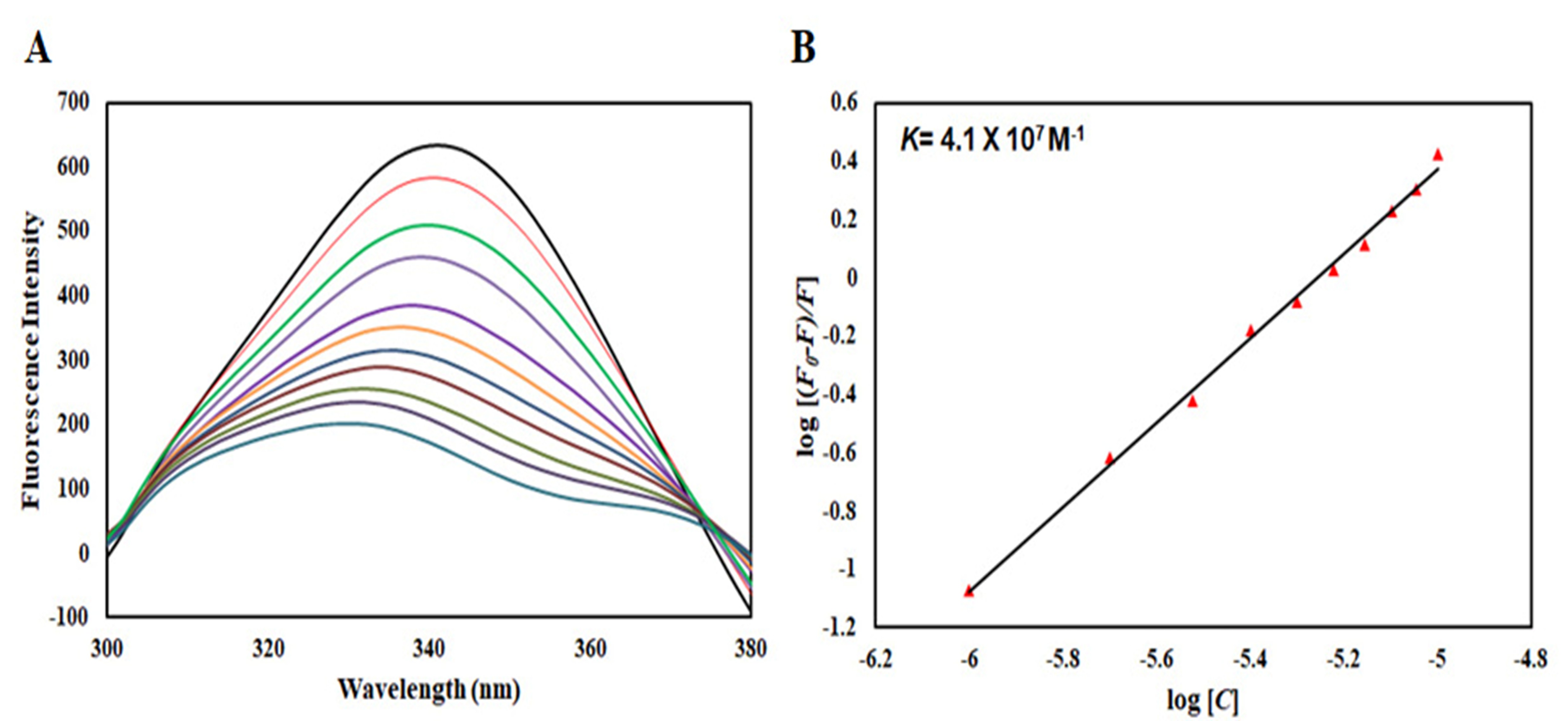
Intrinsic fluorescence spectroscopy was employed to evaluate the interaction between limonene and recombinant CDK6. Progressive addition of limonene led to a marked decrease in the intrinsic fluorescence intensity of CDK6, indicating effective quenching of the protein's fluorescence (Figure 6A). This observation suggests that ligand binding alters the local microenvironment surrounding aromatic fluorophores (primarily tryptophan residues), thereby affecting emission characteristics. The fluorescence quenching data were analyzed using both the Stern–Volmer and modified Stern–Volmer equations to determine the quenching mechanism and binding parameters. The Stern–Volmer plot (Figure 6B) demonstrated a concentration-dependent quenching profile, supporting the formation of a protein–ligand complex.
Binding parameters derived from the modified Stern–Volmer plot (Figure 6B) revealed a binding constant () of , indicative of strong affinity between limonene and CDK6. The slope of the modified plot further provided the number of binding sites (n), suggesting a defined binding interaction. The high binding constant obtained from fluorescence analysis is consistent with the molecular docking results, collectively supporting a stable and favorable interaction between limonene and CDK6.
Figure 6. Fluorescence binding analysis of limonene with CDK6. (A) Intrinsic fluorescence emission spectra of CDK6 recorded in the presence of increasing concentrations of limonene. The excitation wavelength, λex, was fixed at 280 nm, and emission spectra were collected over 300-400 nm. (B) Modified Stern-Volmer plot derived from fluorescence quenching data.
Download: Download full-size image
3.4. Kinase inhibition assay
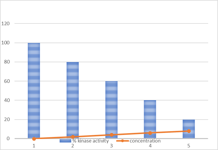
The inhibitory effect of limonene on recombinant CDK6 kinase activity was evaluated in a dose-dependent manner. Increasing concentrations of limonene resulted in a progressive reduction in CDK6 enzymatic activity (Figure 7). The observed decrease in kinase activity demonstrates that limonene effectively suppresses CDK6 function, supporting its potential role as a kinase inhibitor. These findings are consistent with both the molecular docking predictions and fluorescence binding studies, collectively indicating a stable interaction that translates into functional inhibition of the enzyme.
Figure 7. Graph representing enzyme activity versus ligand (limonene) concentration in kinase inhibition assay.
Download: Download full-size image
4. Discussion
CDKs control cell division, which is especially important in cancer. CDK4 and CDK6 are activated by D-cyclins. As a result, CDK4 and CDK6 regulate the G1-to-S transition of the cell cycle. CDK6 plays an important part in the cell cycle. CDK4/6 inhibitors suppress Rb phosphorylation and stop the G1 cell cycle, reducing tumour progression. In many cancers, the CDK4/6–INK4–Rb signalling pathway is dysregulated. CDK4/6 has emerged as a possible therapeutic target (Fontanella et al., 2022). Those CDK4/6 inhibitors may become beneficial for tumour therapy after two decades of exploitation of scientific insights. Cyclin-dependent kinases 4 and 6 (CDK4/6) play a central role in regulating cell cycle progression through the G1–S phase transition, making them critical drivers of uncontrolled proliferation in cancer. Pharmacological inhibition of CDK4/6 has demonstrated potent anti-proliferative effects by inducing cell cycle arrest and promoting senescence in tumor cells. Consequently, CDK4/6 has emerged as a validated therapeutic target across multiple malignancies (Zhu and Zhu, 2023).
Several selective CDK4/6 inhibitors have received clinical approval for the treatment of hormone receptor-positive breast cancer, demonstrating favorable safety profiles and significant therapeutic benefit. Notably,, agents such as Palbociclib, ribociclib, and abemaciclib have substantially improved progression-free survival when administered in combination with endocrine therapy. These findings underscore the clinical value of combination strategies, as CDK4/6 inhibition enhances the efficacy of other therapeutic agents and may help overcome resistance mechanisms (Illia et al., 2026).
Despite these advances, variability in patient response highlights the need for predictive biomarkers to identify individuals most likely to benefit from CDK4/6-targeted therapies. Furthermore, expanding the application of CDK4/6 inhibitors to additional tumor types requires comprehensive preclinical validation and well-designed large-scale clinical trials. Continued research focusing on combination regimens, resistance pathways, and molecular determinants of response will be essential to fully exploit the therapeutic potential of CDK4/6 inhibition in cancer management (Sun et al., 2025).
In this study, we combined computational prediction with biophysical and biochemical validation to investigate limonene as a potential natural inhibitor of CDK6. Molecular docking demonstrated the favorable binding of limonene in the catalytic pocket of CDK6 with a predicted binding free energy of , a value of 4.62, and a ligand efficiency of per non-hydrogen atom. The relatively high ligand efficiency of limonene makes it an effective binding scaffold that could be further optimized through medicinal chemistry approaches to enhance potency while maintaining favorable physicochemical properties. Furthermore, docking analysis showed that limonene occupies the ATP-binding region of CDK6 and establishes stabilizing hydrophobic interactions with residues within the catalytic pocket, with the possibility that it may interfere with ATP binding and can inhibit kinase activity.
After successful expression and purification of recombinant CDK6 with the expected molecular weight checked on SDS–PAGE, the above computational results were further validated experimentally through fluorescence measurements and a kinase inhibition assay. Progressive quenching of CDK6 fluorescence was observed with increasing concentrations of limonene, with the binding constant () of , which indicates the strong binding of limonene and CDK6, supporting the prediction obtained from molecular docking. Functional inhibition of the catalytic activity of CDK6 by limonene was assessed by the malachite green ATPase assay, which confirmed the concentration-dependent reduction in enzyme activity with increasing concentrations of limonene.
Overall, the integration of molecular docking, fluorescence spectroscopy, recombinant protein characterization, and kinase inhibition assays provides complementary evidence supporting the inhibitory potential of limonene. Further validation in cancer cell models and in vivo systems will be necessary to establish its therapeutic potential and facilitate the development of optimized limonene-derived CDK6-targeted anticancer agents.
5. Conclusions
Elevated CDK6 expression has been strongly correlated with the progression and poor prognosis of multiple cancer types, underscoring its importance as a therapeutic target for the development of selective small-molecule inhibitors. Natural compounds continue to represent a valuable reservoir of structurally diverse bioactive molecules with potential applications in oncology. In the present study, limonene was identified as a promising CDK6 inhibitor through integrated in silico, biophysical, and enzymatic analyses. The observed binding affinity and functional suppression of CDK6 activity suggest that limonene may contribute to its anticancer effects, at least in part, by modulating cell cycle regulatory pathways. This mechanistic insight provides a novel perspective on the anticancer potential of limonene. Importantly, the relatively simple chemical scaffold of limonene offers opportunities for rational structural modification to enhance potency, selectivity, and drug-like properties. Structure-guided optimization strategies may facilitate the development of limonene-derived analogs as preclinical leads targeting CDK6. Further cellular and in vivo investigations are required to validate its therapeutic potential and to advance translational applications.
6. Funding
This work received no funding.
7. Acknowledgments
Zulfareen acknowledges The Jamia Millia Islamia for providing a Non-NET fellowship.
8. Conflicts of Interest
The authors declare no conflicts of interest.
9. Data Availability Statement
All data generated or analyzed during this study are included in this manuscript.
10. Declaration on the Use of Artificial Intelligence (AI) Tools
The authors declare that artificial intelligence (AI) tools, specifically ChatGPT (OpenAI), were used solely to refine the language, improve grammar, and enhance the clarity of the manuscript. The AI tool was not used to generate scientific content, analyze data, interpret results, or draw scientific conclusions.
References
O. Bachs, E. Gallastegui, S. Orlando, A. Bigas, J. M. Morante-Redolat, J. Serratosa, I. Farinas. Role of p27Kip1 as a transcriptional regulator. Oncotarget. 9. 26259-26276. 2018. https://doi.org/10.18632/oncotarget.25447
J. E. Bird, J. Marles-Wright, A Giachino. A User's Guide to Golden Gate Cloning Methods and Standards. ACS Synthetic Biology. 11. 3551-3563. 2022. https://doi.org/10.1021/acssynbio.2c00355
M. Classon, E Harlow. The retinoblastoma tumour suppressor in development and cancer. Nature Reviews Cancer. 2. 910-917. 2002. https://doi.org/10.1038/nrc950
S. M. de Figueiredo, N. S. Binda, J. A. Nogueira-Machado, S. A. Vieira-Filho, R. B Caligiorne. The antioxidant properties of organosulfur compounds (sulforaphane). Recent Patents on Endocrine, Metabolic & Immune Drug Discovery. 9. 24-39. 2015. https://doi.org/10.2174/1872214809666150505164138
C. Fontanella, C. A. Giorgi, S. Russo, S. Angelini, L. Nicolardi, T. Giarratano, S. Frezzini, M. Pestrin, D. Palleschi, S. Bolzonello, V. Parolin, E. R. Haspinger, C. De Rossi, F. Greco, L Gerratana. Optimizing CDK4/6 inhibitors in advanced HR+/HER2- breast cancer: A personalized approach. Critical Reviews in Oncology/Hematology. 180. 103848. 2022. https://doi.org/10.1016/j.critrevonc.2022.103848
R. Garzon, G. Marcucci, C. M Croce. Targeting microRNAs in cancer: Rationale, strategies and challenges. Nature Reviews Drug Discovery. 9. 775-789. 2010. https://doi.org/10.1038/nrd3179
S. T. Hallett, M. W. Pastok, R. M. L. Morgan, A. Wittner, K. Blundell, I. Felletar, S. R. Wedge, C. Prodromou, M. E. M. Noble, L. H. Pearl, J. A Endicott. Differential Regulation of G1 CDK Complexes by the Hsp90-Cdc37 Chaperone System. Cell Reports. 21. 1386-1398. 2017. https://doi.org/10.1016/j.celrep.2017.10.042
D. Hanahan, R. A Weinberg. Hallmarks of cancer: The next generation. Cell. 144. 646-674. 2011. https://doi.org/10.1016/j.cell.2011.02.013
N. M. Hassan, A. A. Alhossary, Y. Mu, C. Kwoh. Protein-ligand blind docking using QuickVina-W with inter-process spatio-temporal integration. Scientific Reports. 7. 15451. 2017. https://doi.org/10.1038/s41598-017-15571-7
A. L. Hopkins, C. R. Groom, A Alex. Ligand efficiency: A useful metric for lead selection. Drug Discovery Today. 9. 430-431. 2004. https://doi.org/10.1016/S1359-6446(04)03069-7
M. F. Illia, G. Colucci, A. Rodriguez Mignola, M. Eiben, A. M. Ballester, N. Zeff, F. Paesani, G. Crimi, F. Von Stecher, M. de la Vega, F. Perazzo. BRCA and CDK4/6: Allies or antagonists?-efficacy of CDK4/6 inhibitors in HR(+)/HER2(-) breast cancer with germline BRCA1/2 mutations: A narrative review. Translational Breast Cancer Research. 7. 7. 2026. https://doi.org/10.21037/tbcr-25-46
E. Jameel, H. Naz, P. Khan, M. Tarique, J. Kumar, S. Mumtazuddin, S. Ahamad, A. Islam, F. Ahmad, N Hoda. Design, synthesis, and biological evaluation of pyrimidine derivatives as potential inhibitors of human calcium/calmodulin-dependent protein kinase IV. Chemical Biology & Drug Design. 89. 741-754. 2017. https://doi.org/10.1111/cbdd.12898
J. Kim, D. H. Kim, M. A. Hossain, M. Y. Kim, B. Sung, J. Yoon, H. Suh, T. C. Jeong, H. Y. Chung, N. D Kim. HS-1793, a resveratrol analogue, induces cell cycle arrest and apoptotic cell death in human breast cancer cells. International Journal of Oncology. 44. 473-480. 2013. https://doi.org/10.3892/ijo.2013.2207
Y. Li, J. Zhang, W. Gao, L. Zhang, Y. Pan, S. Zhang, Y Wang. Insights on structural characteristics and ligand binding mechanisms of CDK2. International Journal of Molecular Sciences. 16. 9314-9340. 2015. https://doi.org/10.3390/ijms16059314
M. Malumbres, M Barbacid. Cell cycle, CDKs and cancer: A changing paradigm. Nature Reviews Cancer. 9. 153-166. 2009. https://doi.org/10.1038/nrc2602
M Malumbres. Cyclin-dependent kinases. Genome Biology. 15. 122. 2014. https://doi.org/10.1186/gb4184
T. Mohammad, Y. Mathur, M. I Hassan. InstaDock: A single-click graphical user interface for molecular docking-based virtual high-throughput screening. Briefings in Bioinformatics. 22. bbaa279. 2021. https://doi.org/10.1093/bib/bbaa279
S. Nebenfuehr, K. Kollmann, V Sexl. The role of CDK6 in cancer. International Journal of Cancer. 147. 2988-2995. 2020. https://doi.org/10.1002/ijc.33054
C. C O'Sullivan. Cyclin-Dependent 4/6 Kinase Inhibitors for Treatment of HER2-Positive Breast Cancer: 2026 Update. Cancers. 18. 533. 2026. https://doi.org/10.3390/cancers18030533
G. Petroni, S. C. Formenti, S. Chen-Kiang, L Galluzzi. Immunomodulation by anticancer cell cycle inhibitors. Nature Reviews Immunology. 20. 669-679. 2020. https://doi.org/10.1038/s41577-020-0300-y
O. Rencuzogullari, P. O. Yerlikaya, A. C. Gürkan, E. D. Arisan, D Telci. Palbociclib, a selective CDK4/6 inhibitor, restricts cell survival and epithelial-mesenchymal transition in Panc-1 and MiaPaCa-2 pancreatic cancer cells. Journal of Cellular Biochemistry. 121. 508-523. 2020. https://doi.org/10.1002/jcb.29249
A. A. Russo, L. Tong, J. Lee, P. D. Jeffrey, N. P Pavletich. Structural basis for inhibition of the cyclin-dependent kinase Cdk6 by the tumour suppressor p16INK4a. Nature. 395. 237-243. 1998. https://doi.org/10.1038/26155
G. Sa, T Das. Anti cancer effects of curcumin: Cycle of life and death. Cell Division. 3. 14. 2008. https://doi.org/10.1186/1747-1028-3-14
A. Shah, E. Bloomquist, S. Tang, W. Fu, Y. Bi, Q. Liu, J. Yu, P. Zhao, T. R. Palmby, K. B Goldberg. FDA approval: Ribociclib for the treatment of postmenopausal women with hormone receptor-positive, HER2-negative advanced or metastatic breast cancer. Clinical Cancer Research. 24. 2999-3004. 2018. https://doi.org/10.1158/1078-0432.CCR-17-2369
A. Shamsi, S. Anwar, T. Mohammad, M. F. Alajmi, A. Hussain, M. T. Rehman, G. M. Hasan, A. Islam, M. I Hassan. MARK4 inhibited by AChE inhibitors, donepezil and Rivastigmine tartrate: Insights into Alzheimer's disease therapy. Biomolecules. 10. 789. 2020. https://doi.org/10.3390/biom10050789
C. J. Sherr, D. Beach, G. I Shapiro. Targeting CDK4 and CDK6: From Discovery to Therapy. Cancer Discovery. 6. 353-367. 2016. https://doi.org/10.1158/2159-8290.CD-15-0894
A. Shigenaka, E. Nitta, T. Nakagawa, M. Nakagawa, T Hosoi. Defective Neural Stem and Progenitor Cell Proliferation in Neurodevelopmental Disorders. Journal of Developmental Biology. 13. 40. 2025. https://doi.org/10.3390/jdb13040040
S. Shityakov, C Forster. In silico structure-based screening of versatile P-glycoprotein inhibitors using polynomial empirical scoring functions. Advances and Applications in Bioinformatics and Chemistry. 7. 1-9. 2014. https://doi.org/10.2147/AABC.S56046
T. M. Sielecki, J. F. Boylan, P. A. Benfield, G. L Trainor. Cyclin-dependent kinase inhibitors: Useful targets in cell cycle regulation. Journal of Medicinal Chemistry. 43. 1-18. 2000. https://doi.org/10.1021/jm990426f
L. M. Spring, S. A. Wander, F. Andre, B. Moy, N. C. Turner, A Bardia. Cyclin-dependent kinase 4 and 6 inhibitors for hormone receptor-positive breast cancer: Past, present, and future. The Lancet. 395. 817-827. 2020. https://doi.org/10.1016/S0140-6736(20)30165-3
N. Srivastava, A. K Saxena. Novel Small Molecule Inhibitors of Cyclin-dependent Kinases as Anticancer Agents. Current Medicinal Chemistry. 32. 7512-7553. 2025. https://doi.org/10.2174/0109298673331685241205180307
J Sun. D-Limonene: Safety and clinical applications. Alternative Medicine Review. 12. 2007.
Y. Sun, Y. Zhao, X. Yu, Y. Qi, X Dai. CDK4/6 inhibitor rechallenge therapy in advanced breast cancer. Discover Oncology. 17. 138. 2025. https://doi.org/10.1007/s12672-025-04269-2
A. Tigan, F. Bellutti, K. Kollmann, G. Tebb, V Sexl. CDK6-a review of the past and a glimpse into the future: From cell-cycle control to transcriptional regulation. Oncogene. 35. 3083-3091. 2016. https://doi.org/10.1038/onc.2015.407
O. Trott, A. J Olson. AutoDock Vina: Improving the speed and accuracy of docking with a new scoring function, efficient optimization, and multithreading. Journal of Computational Chemistry. 31. 455-461. 2010. https://doi.org/10.1002/jcc.21334
M. Voura, P. Khan, S. Thysiadis, S. Katsamakas, A. Queen, G. M. Hasan, S. Ali, V. Sarli, M. I Hassan. Probing the inhibition of microtubule affinity regulating kinase 4 by N-substituted acridones. Scientific Reports. 9. 1676. 2019. https://doi.org/10.1038/s41598-018-38217-8
M. Yousuf, A. Shamsi, P. Khan, M. Shahbaaz, M. F. AlAjmi, A. Hussain, G. M. Hassan, A. Islam, Q. M. Rizwanul Haque, M. I Hassan. Ellagic acid controls cell proliferation and induces apoptosis in breast cancer cells via inhibition of cyclin-dependent kinase 6. International Journal of Molecular Sciences. 21. 3526. 2020. https://doi.org/10.3390/ijms21103526
M. Yousuf, M. Alam, A. Shamsi, P. Khan, G. M. Hassan, Q. M. Rizwanul Haque, M. I Hassan. Structure-guided design and development of cyclin-dependent kinase 4/6 inhibitors: A review on therapeutic implications. International Journal of Biological Macromolecules. 218. 394-408. 2022. https://doi.org/10.1016/j.ijbiomac.2022.07.156
Z. Zhu, Q Zhu. Differences in metabolic transport and resistance mechanisms of Abemaciclib, Palbociclib, and Ribociclib. Frontiers in Pharmacology. 14. 1212986. 2023. https://doi.org/10.3389/fphar.2023.1212986