
About Article
Research article • Volume 1, Issue 1, Pages 1-9 (2026) 100001
Genome-wide annotation and structural modeling of hypothetical proteins in Listeria monocytogenes
https://doi.org/10.53295/cmb.v1.i1.100001
Under a Creative Commons license
Open access
Abstract
The rapid expansion of genome sequencing projects has resulted in the identification of numerous hypothetical proteins whose functions remain uncharacterized. In Listeria monocytogenes serotype 4b, a major food-borne pathogen associated with high mortality rates, several predicted proteins lack functional annotation despite their potential role in pathogenicity and survival. In the present study, a comprehensive in silico approach was employed to functionally annotate 92 hypothetical proteins identified from the genome of L. monocytogenes. Protein sequences were retrieved and analyzed using sequence similarity searches, conserved domain identification, motif analysis, and multiple sequence alignment. Functional classification was performed based on BLAST, Pfam, and InterPro analyses. Structural prediction was performed via homology modeling and the SWISS-MODEL server, followed by structural validation and comparative analysis using PyMOL and DALI-Lite. Functional inference from structural models was supported by conserved-residue mapping and ProFunc analysis. Sequence-based analysis enabled classification of most hypothetical proteins into functional groups, including hydrolases, transferases, transporters, kinases, stress-response proteins, membrane proteins, DNA-binding proteins, and ATP-binding proteins. Structure-based modeling of selected proteins further confirmed the predicted catalytic residues, metal-binding sites, ligand-interaction sites, and conserved functional motifs. Several proteins were predicted to be involved in enzymatic activity, nucleotide metabolism, membrane transport, transcriptional regulation, and stress adaptation. This integrative computational analysis provides functional insights into previously uncharacterized proteins of L. monocytogenes. The findings enhance genome annotation quality and identify potential targets for further experimental validation, contributing to a better understanding of bacterial physiology and pathogenesis.
Keywords: Listeria monocytogenes, hypothetical proteins, functional annotation, homology modeling, genome analysis, structural prediction, protein function prediction
Introduction
Due to the speed and cost-effectiveness of genome sequencing, many small bacterial, archaeal, and eukaryotic genomes have been sequenced, and larger eukaryotic genomes are expected to be fully sequenced soon. In such a case, annotation becomes problematic when genes are predicted faster than annotation can keep pace. Despite ongoing improvements in genome annotation, a substantial proportion of open reading frames remain classified as "conserved hypothetical proteins" (Gury et al., 2004). Hypothetical proteins are predicted from nucleotide sequences but lack direct experimental validation at the protein level. In many instances, these predicted proteins show limited sequence similarity to previously characterized and annotated proteins. Conserved hypothetical proteins constitute a significant fraction of genes in sequenced genomes; they encode proteins identified across multiple phylogenetic lineages but remain uncharacterized with respect to biological function or biochemical properties, sometimes accounting for a large portion of the predicted proteome (Sivashankari, 2006).
Determining the functions of hypothetical proteins is essential for improving genomic and proteomic annotations. The identification of new hypothetical proteins can reveal previously unknown structural folds and biological activities (Lubec et al., 2005). As more structures are characterized, novel domains, motifs, and conformational arrangements are likely to emerge, contributing to a clearer understanding of structure–function relationships in proteins. Functional characterization may also uncover new molecular pathways and regulatory cascades. Furthermore, hypothetical proteins may serve as biomarkers or therapeutic targets. Their identification can facilitate the discovery of previously unrecognized or computationally predicted genes. Ultimately, elucidating the roles of these proteins will deepen our understanding of biological systems and may contribute to the development of improved therapeutic interventions.
The biological roles of many predicted proteins remain unknown because their existence has been inferred solely from genomic sequences. The rapid completion of genome sequencing projects has generated extensive biological datasets, enabling the association of genes with their corresponding protein products, which are fundamental to cellular function. Advances in genome and cDNA sequencing technologies allow the prediction of numerous proteins for which experimental evidence is lacking. Functional insights into hypothetical proteins are often derived from sequence comparisons with proteins of known function in model organisms. Since similarities in sequence or conserved domains frequently indicate shared functional properties, such analyses can suggest potential biological roles. Moreover, if a hypothetical protein exhibits significant structural homology with characterized proteins, it is reasonable to infer comparable molecular features.
Traditional biochemical and molecular biology approaches can accurately assign functions to genes; however, these experimental methods are labor-intensive and costly. Consequently, even in fully sequenced genomes, only about 50–60% of genes have been functionally annotated. Automated genome analysis and computational annotation strategies provide alternative means to interpret genomic information. Therefore, determining protein function remains a major challenge in the post-genomic era. This has increased reliance on bioinformatics tools to predict the roles of uncharacterized protein sequences, and several contemporary computational approaches have been developed to address this issue.
Listeria monocytogenes (L. monocytogenes) is the causative agent of listeriosis and is a facultative anaerobic bacterium. The organism can invade and proliferate within host cells and is recognized as one of the most severe foodborne pathogens, with mortality rates of 20–30% in clinical cases (Gebremedhin et al., 2021). In the United States, listeriosis is responsible for approximately 2,500 reported cases and 500 deaths annually, making it one of the leading causes of death among foodborne bacterial infections (Gebremedhin et al., 2021). Although relatively rare, the disease is associated with a high case-fatality rate. Its emergence as a public health concern has been attributed to changing dietary habits, advances in food preservation technologies that extend shelf life, and the bacterium's ability to survive and grow at refrigeration temperatures.
Listeriosis is a bacterial infection caused by L. monocytogenes. It primarily affects individuals at increased risk, including pregnant women, newborns, people with diabetes, cancer, or kidney disease, individuals with AIDS, and those receiving glucocorticosteroid therapy. L. monocytogenes causes flu-like symptoms and meningitis if it spreads to the central nervous system. The first reported case of listeriosis was documented in 1981. Since then, numerous listeriosis outbreaks have been detected and investigated. Subsequent studies have confirmed that serotype 4b is one of the major serotypes associated with human listeriosis. Of the 13 recognized serotypes of L. monocytogenes, serotype 4b is among the most prominent and is frequently associated with invasive infections. L. monocytogenes infection during pregnancy can result in miscarriage, stillbirth, or preterm birth, particularly among women with underlying health conditions. The risk of adverse fetal outcomes is increased when the mother is infected with L. monocytogenes (Nuesch-Inderbinen et al., 2021).
The L. monocytogenes genome is approximately 3 Mb in size and consists of a single chromosome. The reference genome contains 2,912,690 bp, 2,766 predicted protein-coding genes, and 85 RNA genes. Under stressful conditions, L. monocytogenes expresses several stress-response proteins that contribute to its ability to survive in harsh environments. Genome analysis of L. monocytogenes serotype 4b has identified 92 hypothetical proteins, for which limited functional information is currently available. Here, our aim is to annotate the hypothetical proteins from Listeria monocytogenes by using recent bioinformatics tools. After annotating a genome, we grouped genes into two types: the first comprises known genes with functional characterization; the second comprises conserved hypothetical genes conserved across many organisms. Generally, a newly sequenced genome contains approximately 30% of genes that are poorly or incorrectly annotated (Gury et al., 2004). Hence, these poorly characterized hypothetical genes might play an important role in our understanding of the pathogen's pathogenesis and virulence. Given that so little is known about these genes, we have attempted to assign plausible functions to hypothetical proteins. Functional evidence suggests that these hypothetical proteins may have specific functions; however, further detailed analysis is required to confirm their functions.
2. Materials and Methods
2.1. Data mining
We have analyzed the L. monocytogenes genome and its hypothetical proteins by retrieving relevant information from the Protein Information Resource (PIR) database. We have identified and downloaded 92 hypothetical protein sequences of L. monocytogenes. The analyzed protein sequences, together with their primary accession numbers, lengths, pI values, and proposed functions, are presented in Table 1. Protein sequences are saved in FASTA format for further processing. A systematic pipeline used in this study is illustrated in Figure 1.
Figure 1. Outline of structure and function assignment to the hypothetical proteins.
Download: Download full-size image
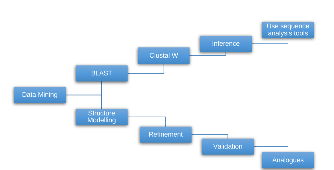
Table 1. List of hypothetical proteins in \textit{Listeria monocytogenes}.
| S. No. | Name of Hypothetical Protein | pI | Length | Pfam | Proposed Function |
|---|---|---|---|---|---|
| 1 | C1L300/ C1L300_LISMC | 9.69 | 447 | MatE | (PF01554) antiporter activity, drug transmembrane transporter activity |
| 2 | C1L2X5/ C1L2X5_LISMC | 7.85 | 345 | UPF0118 | (PF01594) NA |
| 3 | C1L2V8/ C1L2V8_LISMC | 5.02 | 120 | DUF964 | (PF06133) NA |
| 4 | C1L2V7/ C1L2V7_LISMC | 5.45 | 267 | YmdB | (PF13277) putative phosphoesterases |
| 5 | C1L2S2/ C1L2S2_LISMC | 6.41 | 274 | FtsJ | (PF01728) S4 (PF01479) methyltransferase involved in viral RNA capping, RNA |
| 6 | C1L2Q9/ C1L2Q9_LISMC | 6.47 | 321 | DUF1385 | (PF07136) NA |
| 7 | C1L2P0/ C1L2P0_LISMC | 4.78 | 118 | No domains | NA |
| 8 | C1L2N2/ C1L2N2_LISMC | 5.44 | 92 | DUF503 | (PF04456) NA |
| 9 | C1L2K8/ C1L2K8_LISMC | 5.38 | 174 | Acetyltransf_3 | (PF13302) transfers acetyl group |
| 10 | C1L2J0/ C1L2J0_LISMC | 7.84 | 103 | NA | NA |
| 11 | C1L2E5/ C1L2E5_LISMC | 5.13 | 174 | Metallophos_2 | (PF12850) Phosphoesterase (hydrolase activity, acting on ester bonds) |
| 12 | C1L1Z7/ C1L1Z7_LISMC | 6.42 | 774 | Glycos_transf_2 | Glycos_transf_2 (PF00535); Glyphos_transf (PF04464) |
| 13 | C1L1V8/ C1L1V8_LISMC | 9.43 | 267 | DUF817 | (PF05675) NA |
| 14 | C1L1U8/ C1L1U8_LISMC | 6.18 | 555 | Lactamase_B | Lactamase_B (PF00753); RNA binding; hydrolase activity, acting on ester bonds, metal-dependent |
| 15 | C1L1S5/ C1L1S5_LISMC | 10.1 | 344 | DUF218 | (PF02698) NA |
| 16 | C1L1R3/ C1L1R3_LISMC | 9.3 | 255 | TerC | (PF03741) integral to membrane |
| 17 | C1L1R2/ C1L1R2_LISMC | 9.36 | 255 | TerC | (PF03741) integral to membrane |
| 18 | C1L1R1/ C1L1R1_LISMC | 9.09 | 453 | MatE | (PF01554) antiporter activity, drug transmembrane transporter activity |
| 19 | C1L1K4/ C1L1K4_LISMC | 9.81 | 201 | SNARE_assoc | (PF09335) NA |
| 20 | C1L1G8/ C1L1G8_LISMC | 5.59 | 725 | Tex_N | (PF09371), HHH_3 (PF12836), S1 RNA binding, hydrolase activity- acting on ester bonds |
| 21 | C1L1B3/ C1L1B3_LISMC | 4.78 | 125 | Ribonuc_L-PSP | (PF01042) Inhibits protein synthesis by cleaving the mRNA |
| 22 | C1L190/ C1L190_LISMC | 5.09 | 282 | NA | NA |
| 23 | C1L165/ C1L165_LISMC | 4.69 | 176 | YceI | (PF04264) binds to polyisoprenoid |
| 24 | C1L164/ C1L164_LISMC | 9.54 | 306 | EamA | (PF00892) transporter activity |
| 25 | C1L162/ C1L162_LISMC | 9.27 | 233 | DUF554 | (PF04474) NA |
| 26 | C1L161/ C1L161_LISMC | 4.79 | 296 | CN_hydrolase | (PF00795) hydrolase activity, acting on carbon-nitrogen (but not peptide) |
| 27 | C1L158/ C1L158_LISMC | 5.68 | 282 | PhzC-PhzF | (PF02567) Phenazine biosynthes |
| 28 | C1L143/ C1L143_LISMC | 5.33 | 309 | DAGK_cat | (PF00781) NAD+ kinase activity, diacylglycerol kinase activity |
| 29 | C1L0U3/ C1L0U3_LISMC | 4.83 | 288 | Hydrolase_3 | (PF08282) hydrolase activity |
| 30 | C1L0T5/ C1L0T5_LISMC | 5.42 | 209 | HhH-GPD | (PF00730) DNA binding, endonuclease activity |
| 31 | C1L0T0/ C1L0T0_LISMC | 9.82 | 187 | DUF420 | (PF04238) NA |
| 32 | C1L0R8/ C1L0R8_LISMC | 5.34 | 606 | Sulfatase | (PF00884) sulfuric ester hydrolase activity |
| 33 | C1L0Q0/ C1L0Q0_LISMC | 9.71 | 246 | TauE | (PF01925) NA |
| 34 | C1L0P3/ C1L0P3_LISMC | 4.87 | 172 | Acetyltransf_1 | (PF00583) acetyltransferase activity |
| 35 | C1L0M8/ C1L0M8_LISMC | 4.78 | 110 | PadR | (PF03551) involved in negative regulation of phenolic acid metabolism |
| 36 | C1L0L0/ C1L0L0_LISMC | 6.08 | 394 | Methyltrans_SAM | (PF10672) methyltransferase activity |
| 37 | C1L0K2/ C1L0K2_LISMC | 7.7 | 431 | Xan_ur_permease | (PF00860) transporter activity |
| 38 | C1L0G9/ C1L0G9_LISMC | 4.82 | 264 | AP_endonuc_2 | (PF01261) involved in the myo-inositol catabolism (isomerase activity) |
| 39 | C1L075/ C1L075_LISMC | 5.18 | 346 | Lactonase | (PF10282) 6-phosphogluconolactonase activity |
| 40 | C1L028/ C1L028_LISMC | 5.72 | 143 | Usp | (PF00582) response to stress |
| 41 | C1L024/ C1L024_LISMC | 5.26 | 226 | GATase | (PF00117) transferase activity |
| 42 | C1KZV6/ C1KZV6_LISMC | 4.89 | 281 | NmrA | (PF05368) nucleotide binding (negative transcriptional regulator |
| 43 | C1KZU1/ C1KZU1_LISMC | 5.41 | 377 | Gly_kinase | (PF02595) glycerate kinase activity |
| 44 | C1KZQ3/ C1KZQ3_LISMC | 4.64 | 279 | Hydrolase_3 | (PF08282) hydrolase activity |
| 45 | C1KZM7/ C1KZM7_LISMC | 8.93 | 156 | Usp | (PF00582) response to stress |
| 46 | C1KZM4/ C1KZM4_LISMC | 6.15 | 120 | YjbR | (PF04237) NA |
| 47 | C1KZE1/ C1KZE1_LISMC | 4.83 | 270 | Hydrolase_3 | (PF08282) hydrolase activity |
| 48 | C1KZ86/ C1KZ86_LISMC | 5.18 | 111 | PhnA_Zn_Ribbon | (PF08274), PhnA NA |
| 49 | C1KZ81/ C1KZ81_LISMC | 5.97 | 494 | FTR1 | (PF03239) transmembrane transport |
| 50 | C1KZ17/ C1KZ17_LISMC | 8.78 | 220 | MgtC | (PF02308) transport of Mg2+ |
| 51 | C1KZ02/ C1KZ02_LISMC | 5.31 | 280 | SAM_adeno_trans | (PF01887) NA |
| 52 | C1KYZ6/ C1KYZ6_LISMC | 8.7 | 362 | MacB_PCD | (PF12704), FtsX (PF02687) transport lipids targeted to the outer membrane across the |
| 53 | C1KYZ4/ C1KYZ4_LISMC | 5.44 | 97 | ABM | (PF03992) monooxygenase activity |
| 54 | C1KYZ2/ C1KYZ2_LISMC | 4.62 | 275 | Hydrolase_3 | (PF08282) hydrolase activity |
| 55 | C1KYY1/ C1KYY1_LISMC | 5.4 | 440 | HD | (PF01966) metal ion binding, phosphoric diester hydrolase activity |
| 56 | C1KYX9/ C1KYX9_LISMC | 6.83 | 217 | Peptidase_M50 | (PF02163) proteolysis, metalloendopeptidase activity |
| 57 | C1KYX3/ C1KYX3_LISMC | 9.03 | 306 | DAGK_cat | (PF00781) kinase, transferase |
| 58 | C1KYW9/ C1KYW9_LISMC | 10.1 | 357 | UPF0104 | (PF03706) NA |
| 59 | C1KYT3/ C1KYT3_LISMC | 5.78 | 211 | UPF0029 | (PF01205), DUF1949 (PF09186) GTP binding |
| 60 | C1KYS5/ C1KYS5_LISMC | 6.43 | 287 | DUF161 | (PF02588), DUF2179 (PF10035) NA |
| 61 | C1KYP0/ C1KYP0_LISMC | 4.97 | 322 | UPF0052 | (PF01933) NA |
| 62 | C1KYM2/ C1KYM2_LISMC | 5.12 | 259 | CN_hydrolase | (PF00795) hydrolase activity, acting on carbon-nitrogen (but not peptide) |
| 63 | C1KYL6/ C1KYL6_LISMC | 5.39 | 273 | Hydrolase_3 | (PF08282) ATP binding, ATPase activity, coupled to transmembrane |
| 64 | C1KYI4/ C1KYI4_LISMC | 5.98 | 201 | MTS | (PF05175) 16S rRNA (guanine(966)-N(2))-methyltransferase activity |
| 65 | C1KY64/ C1KY64_LISMC | 5.76 | 117 | ArsC | (PF03960) regulate the transcription of multiple genes in response to |
| 66 | C1KY61/ C1KY61_LISMC | 5.27 | 291 | Cation_efflux | (PF01545) cation transmembrane transporter activity |
| 67 | C1KY51/ C1KY51_LISMC | 5.94 | 147 | NifU_N | (PF01592) iron ion binding, iron-sulfur cluster binding |
| 68 | C1KY50/ C1KY50_LISMC | 4.87 | 464 | UPF0051 | (PF01458) iron-sulfur cluster assembly |
| 69 | C1KY46/ C1KY46_LISMC | 9.02 | 279 | TauE | (PF01925) involved in the transport of anions across the cytoplasmic |
| 70 | C1KY45/ C1KY45_LISMC | 5.08 | 462 | Metallophos | (PF00149), 5_nucleotid_C hydrolase activity (5_nucleotid_C) |
| 71 | C1KY43/ C1KY43_LISMC | 4.94 | 255 | Hydrolase_like | (PF13242), Hydrolase_6 hydrolase activity |
| 72 | C1KY41/ C1KY41_LISMC | 4.82 | 436 | DUF21 | (PF01595), CBS (PF00571), flavin adenine dinucleotide binding (CBS), |
| 73 | C1KY30/ C1KY30_LISMC | 9.43 | 406 | Voltage_CLC | (PF00654) voltage-gated chloride channel activity |
| 74 | C1KY03/ C1KY03_LISMC | 5.29 | 156 | Rrf2 | (PF02082) NA |
| 75 | C1KXZ5/ C1KXZ5_LISMC | 4.65 | 281 | Hydrolase_3 | (PF08282) hydrolase activity |
| 76 | C1KXZ0/ C1KXZ0_LISMC | 4.76 | 532 | Amidohydro_3 | (PF07969) hydrolase activity, acting on carbon-nitrogen (but not peptide) |
| 77 | C1KXY4/ C1KXY4_LISMC | 6.26 | 257 | Methyltransf_26 | (PF13659) methyltransferase activity |
| 78 | C1KXX7/ C1KXX7_LISMC | 4.83 | 270 | Hydrolase_3 | (PF08282) hydrolase activity |
| 79 | C1KXV5/ C1KXV5_LISMC | 5.52 | 249 | EAL | (PF00563) NA |
| 80 | C1KXN7/ C1KXN7_LISMC | 6.53 | 331 | Bac_luciferase | (PF00296) oxidoreductase activity, acting on paired donors, with |
| 81 | C1KXN1/ C1KXN1_LISMC | 6.58 | 147 | DUF523 | (PF04463) NA |
| 82 | C1KWJ0/ C1KWJ0_LISMC | 6.74 | 121 | DUF1798 | (PF08807) NA |
| 83 | C1KWG8/ C1KWG8_LISMC | 6.52 | 327 | DUF939 | (PF06081), DUF939_C NA |
| 84 | C1KWG7/ C1KWG7_LISMC | 4.39 | 125 | Glyoxalase_2 | (PF12681) NA |
| 85 | C1KWG4/ C1KWG4_LISMC | 6.52 | 205 | HTH_11 | (PF08279), CBS (PF00571) sequence-specific DNA binding transcription factor activity |
| 86 | C1KWC7/ C1KWC7_LISMC | 5.29 | 291 | YicC_N | (PF03755), DUF1732 (PF08340) play a role in the stationary phase survival (YicC), |
| 87 | C1KW03/ C1KW03_LISMC | 6.11 | 126 | OsmC | (PF02566) has a novel pattern of oxidative stress regulation |
| 88 | C1KVW1/ C1KVW1_LISMC | 6.22 | 192 | rRNA_methylase | (PF06962) methyltransferase activity |
| 89 | C1KVW0/ C1KVW0_LISMC | 5.48 | 321 | Radical_SAM | (PF04055) catalytic activity, iron-sulfur cluster binding |
| 90 | C1KVA0/ C1KVA0_LISMC | 6.02 | 234 | DUF633 | (PF04816) tRNA (adenine-N1-)-methyltransferase activity |
| 91 | C1KV99/ C1KV99_LISMC | 5.5 | 373 | NIF3 | (PF01784) NIF3 interacts with the yeast transcriptional coactivator |
| 92 | C1KV71/ C1KV71_LISMC | 4.92 | 269 | Hydrolase_3 | (PF08282) cation transport, ATP binding, ATPase activity, coupled to |
2.2. Sequence similarity search
The functions of the hypothetical proteins were inferred based on sequence similarity to well-characterized homologs in GenBank. Protein-protein BLAST (BLASTP) was used to compare the query amino acid sequences with sequences in protein databases and identify homologous hits. BLASTP results against public databases provided insights into the potential functional properties of the related proteins.
2.3. Structure Prediction
The three-dimensional structures of hypothetical proteins were examined to infer their potential biological functions. Structural prediction was carried out using homology modeling, which generally involves multiple sequential steps. First, a suitable template protein with an experimentally determined structure, typically belonging to the same or a closely related family, is identified. Second, appropriate sequence alignment tools are employed to obtain the best possible alignment between the target and template proteins. Third, the target sequence is divided into smaller segments to facilitate accurate modeling. Fourth, corresponding fragments are searched in structural databases based on sequence similarity and the spatial conformation of the selected template. Fifth, the spatial coordinates of these matched segments are assembled and adjusted to construct the full-length target structure, ensuring that all atomic positions are properly incorporated. These procedures are repeated multiple times to generate several models, from which an averaged structure is obtained, followed by comprehensive energy minimization to refine the final predicted model. The 3D structures were retrieved using the SWISS-MODEL server, an automated comparative modeling system for protein structure prediction. The homology modeling workflow was performed in the project mode of PyMOL, an integrated sequence-structure workbench. Using the SWISS-MODEL server, we obtained suitable 3D structures for thirteen hypothetical proteins selected for detailed structural analysis, and the resulting structural models were downloaded in PDB format for further analysis.
2.4. Function prediction
Clustering of gene expression profiles is widely used for functional prediction, based on the principle that genes involved in related biological processes tend to exhibit coordinated expression patterns. Functional inference can also be derived from protein–protein interaction networks, where the role of an uncharacterized protein is estimated from the known functions of its interacting partners. Schwikowski and co-workers introduced a neighbor-counting strategy in which the probable function of an unknown protein is determined according to the frequency of functional annotations among its interaction partners. Hishigaki et al., 2001 through the incorporation of a chi-square (χ^2) statistical framework to enhance prediction reliability. In both methods, all functional contributions from neighboring proteins are treated with equal significance. Several publicly accessible signature and motif databases support functional annotation and are frequently integrated with sequence clustering and domain-based resources. These include PROSITE, PRINTS, Pfam, ProDom, Blocks, SMART, and InterPro; in the present study, Pfam and InterPro were specifically utilized. Structural classification of proteins into homologous families, superfamilies, and folds can be performed using databases such as SCOP, CATH, FSSP, CAMPASS, and HOMSTRAD. Additionally, the ProFunc server facilitates structure-based functional prediction by comparing modeled protein structures against multiple databases and reporting significant matches for functional interpretation.
2.5. Visualization
We carried out structural analysis using PyMOL for superposition of molecules, secondary-structure-based alignment, and calculation of RMSD values. PyMOL is also used to depict structures, as well as to analyze potential residue positions and functions. We visualized the template residues and the model after superimposing the model onto the template. Each residue was visualized individually, and residue conservation was assessed.
2.6. Structure analysis
Structure-based comparison approaches are highly effective for detecting remote evolutionary relationships that may not be apparent from sequence similarity alone. Structural information also enables the recognition of functional sites that have arisen independently during evolution. Because protein function is inherently dependent on three-dimensional conformation, structural analysis provides direct insight into the molecular mechanisms underlying biological activity. Local structural alignment focuses on identifying conserved three-dimensional arrangements of specific residues, even among proteins that differ substantially in overall fold. Several computational tools are available for such analyses, including DaliLite, which utilizes C–C distance matrix comparisons; TOPS; SSM; VAST; GRATH, which represents secondary structure elements as graph nodes connected by spatial relationships; and SSAP, which evaluates structural similarity based on C comparisons and additional scoring features. Other methods such as LSQMAN, CE (Combinatorial Extension), MATRAS, and LOCK2 also employ atom-based or secondary structure vector representations for alignment. In the present study, DALI-Light was selected for structural comparison and analysis.
3. Results and Discussion
3.1. Sequence-based Function Prediction
Information derived from BLAST, ClustalW, and other online servers for sequence analysis revealed close similarities of hypothetical proteins to proteins of known function. Based on the proposed functions, we classified all 92 hypothetical proteins into functional classes. All the Hypothetical proteins have been classified into the following categories.
Hydrolase. The genomic proteins of L. monocytogenes showed the greatest similarity to proteins with hydrolase activity. Based on comparisons of conserved residues and sequence similarity with known characterized proteins, C1L0U3, C1L0R8, C1KZQ3, C1KZE1, C1KYZ2, C1KYY1, C1KY45, C1KY43, and C1KXZ5 were predicted to possess hydrolase activity. Hydrolases are enzymes that facilitate hydrolytic reactions, in which a water molecule is utilized to break chemical bonds within a substrate. During this process, the hydrogen (H^+) and hydroxyl (OH^-) components of water are incorporated into the reacting molecule, resulting in its cleavage into two or more smaller products. The term "hydrolase" serves as the systematic designation for enzymes classified under Enzyme Commission (EC) class 3, which encompasses all enzymes that catalyze hydrolysis reactions. For the C1L0U3 protein, 65 matching sequences were identified by FASTA search. Eight neighbouring proteins—Q8Y970, D2PAB8, D2NZC4, C1L0U3, E1UE28, B8DIC3, Q92DZ3, and D3UKJ4—showed sequence identities ranging from 91.5% to 100%. Ten sequence motifs were identified through a search against the InterPro database. For the C1KZQ3 protein, 57 matching sequences were identified using FASTA, while seven neighbouring proteins—E1UBT1, B8DAU2, Q8Y3Y0, D2P7B6, D2NVU7, Q927E2, and D3USV7—showed sequence identities ranging from 83.5% to 99.3%. Nine sequence motifs were identified using the InterPro database. For the C1KY43 protein, 47 matching sequences were identified through a FASTA search, and nine sequence motifs were identified using InterProScan. Seven neighbouring proteins—E1UAS5, B8DDF3, Q8Y4N4, D2P959, D2NY72, Q928N2, and D3UR85—showed sequence identities ranging from 94.0% to 98.0%.
Transferase. Transferases are enzymes that catalyze the movement of specific chemical groups, such as methyl, glycosyl, acyl, or phosphate groups, from one molecule to another. In these reactions, one compound acts as the donor of the functional group, while another serves as the acceptor. The nomenclature and classification of transferases follow the format "donor:acceptor group transferase," reflecting the nature of the transferred group and the participating substrates. Proteins belonging to this group are C1L2K8, C1L1Z7, C1L0P3, C1L0L0, C1L024, C1KYI4, C1KXY4, C1KVW1, and C1KVA0. The protein sequence has been found to be similar to that of characterized proteins in this category. This group is the second most abundant in Listeria monocytogenes, after hydrolases. For the gene C1L0L0, 65 matching sequences were identified by a FASTA search, and 8 motifs were matched by InterProScan, which searches the InterPro database. The 7 neighbouring genes are as follows D2PA32, D2NYL3, Q8Y9E8, E1UDU6, B8DA44, Q92E70 and D3USH9 with percentage identity as 99.5%, 99.5%, 99.5%, 96.7%, 96.7%, 92.8% and 89.4% respectively. There are 29 matching sequences found by FASTA search for the C1KXY4 gene and 6 motifs matched while running it against InterPro database. There are 6 neighboring genes found which are E1UCN5, B8DGQ6, Q8YAG1, D2PAN3, D2NZA9 and Q92F98 with percentage identity as 97.1%, 97.1%, 95.5%, 95.1%, 95.1% and 95.9% respectively.
Transporter. Transporter proteins are proteins that move substances within an organism. Transport proteins are vital to the growth and life of all living things. There are several different kinds of transport proteins. The proteins found in Listeria monocytogenes included in this category are C1L300, C1L1R1, C1L164, C1L0K2, C1KZ81, C1KZ17, C1KYZ6, C1KY61, and C1KY46. For the C1L1R1 gene, 4 matching sequences were found by FASTA search, and 6 motifs were found by InterPro scan. There were 7 neighboring genes found which are E1UF08, B8DEE5, Q8Y8B8, D2P3J7, D2P0R1, Q92D31, and D3ULQ9 with percentage identity as follows 97.9%, 97.9%, 96.8%, 96.6%, 96.6%, 93.4%, and 87.4%, respectively.
RNA Binding. The following residues belong to this group: C1L2S2, C1L1U8, and C1L1G8. RNA-binding proteins are proteins that bind to RNA recognition motifs (RRMs) of double or single-stranded RNA in cells and participate in forming ribonucleoprotein complexes. They are cytoplasmic and nuclear proteins. For the C1L2S2 gene, 15 matching sequences were identified by FASTA search, and 11 sequence motifs were identified in InterPro. The 7 neighbouring genes are Q8Y7C0, D2P4S6, D2P1Z0, E1UG75, B8DFW2, Q92BY9 and D3UMV0 with sequence identity as 97.4%, 97.4%, 97.4%, 95.2%, 95.2%, 94.9% and 94.9% respectively. Whereas for the C1L1U8 gene, 51 matching sequences were found, and 11 sequence motifs matched in InterPro. There are 7 neighbouring genes which are as follows Q92CZ5, Q7AP12, E1UF45, D2P3N4, D2P0U8, B8DEA8 and D3ULU6 with similarities as 100%, 100%, 100%, 100%, 100%, 100%, and 99.5% respectively.
Hydrolase acting on carbon-nitrogen but not on peptide. Following mentioned are the proteins C1L161, C1KYM2, and C1KXZ0, which belong to this category. This family contains hydrolases that break carbon-nitrogen bonds. This sub family of hydrolase is numbered as EC 3.5. There were 35 matching sequences found by FASTA search and 6 sequence motifs matched in the InterPro scan. There were 6 neighbouring genes found which are E1UD07, D2P8J8, D2NWP2, B8DEW3, Q8YA77, and Q92EZ8 with percentage identity as 99.2%, 99.2%, 99.2%, 99.2%, 98.1% and 95.3% respectively.
Kinase. Kinases are enzymes that catalyze the transfer of phosphate groups from high-energy donor molecules, typically adenosine triphosphate (ATP), to specific target substrates in a reaction known as phosphorylation. Through this process, kinases regulate numerous cellular activities, including signal transduction, metabolism, and cell cycle progression. They belong to the broader class of phosphotransferases. In the present study, proteins C1L143, C1KZU1, and C1KYX3 were identified as members of this enzyme category.
Response to stress. Cells exposed to stressful conditions often accumulate abnormal, misfolded, or damaged proteins as a consequence of environmental or physiological challenges. To mitigate these effects, cells increase the production of stress-related proteins that assist in stabilizing, refolding, or degrading altered proteins. Rather than solely enabling survival under extreme or lethal stress, these stress proteins also play a crucial role in cellular recovery following stress exposure, thereby helping restore normal cellular homeostasis. C1LO28, C1KZM7, and C1KW03 are the proteins that belong to this category. A FASTA search of the C1KZM7 gene identified 21 matching sequences, and an InterPro scan identified 4 sequence motifs. Q927G8, E1UBQ4, D2P7S2, D2NVX3, B8DAW9, Q8Y405 and D3USS9 are the 7 neighbouring genes found with 99.3%, 99.3%, 99.3%, 99.3%, 99.3%, 98.6% and 98.6% identity respectively.
Integral to membrane. An integral membrane protein is a protein molecule, or protein complex, that is permanently embedded within the plasma membrane through hydrophobic regions that interact strongly with the lipid bilayer. These proteins are tightly associated with membrane phospholipids and cannot be easily removed without disrupting the membrane structure. Integral membrane proteins are broadly divided into two principal categories: (1) transmembrane proteins and (2) integral monotropic proteins. Transmembrane proteins represent the more prevalent group and extend completely across the lipid bilayer. Such proteins perform diverse biological functions, including acting as transporters, ion channels, receptors, enzymes, structural anchors, components involved in energy storage and signal transduction, and mediators of cell–cell adhesion. Representative examples include integrins, cadherins, the insulin receptor, neural cell adhesion molecules (NCAM), selectins, glycophorin, and rhodopsin. C1L1R3, C1L1R2, and C1KY64 are the proteins that belong to this group. For the gene C1KY64, we found 39 matching sequences by FASTA search and 5 motifs matched in InterPro scan. 6 neighbouring genes found were E1UAU6, B8DDD2, Q8Y4L1, D2P980, D2NY93, and Q928L2 with percentage identity as 98.3%, 98.3%, 96.6%, 96.6%, 96.6%, and 97.4%, respectively.
DNA Binding. DNA-binding proteins are proteins that contain a DNA-binding domain, which in turn is responsible for binding to single or double-stranded DNA molecules. C1L0T5 and C1KWG4 are the proteins that have this domain and are therefore classified in this category.
Phosphoesterase. Phosphoesterases are a family of hydrolases that catalyse the hydrolysis of phosphoester bonds. The proteins C1L2V7 and C1L2E5 were classified as phosphoesterases. For the C1L2E5 protein, 24 matching sequences were identified by FASTA search, and six sequence motifs were identified using InterPro. Seven neighbouring proteins—Q8Y7N4, E1UFM2, B8DHZ2, D2P485, D2P1E9, Q92CG9, and D3UME0—showed sequence identities of 100%, 100%, 100%, 99.4%, 99.4%, 97.6%, and 95.3%, respectively.
ATP Binding. An ATP-binding protein is a protein that specifically interacts with adenosine 5^-triphosphate (ATP), a ribonucleotide composed of the purine base adenine attached to the sugar D-ribofuranose and linked to three phosphate groups. ATP serves as a primary carrier of chemical energy and phosphate groups within the cell. Proteins that bind ATP typically utilize this interaction to drive biochemical reactions, regulate cellular processes, or facilitate molecular transport, thereby playing essential roles in energy-dependent cellular functions. C1KYL6 and C1KV71 are the proteins in this category. For the C1KYL6 gene, we identified 69 matching sequences via a FASTA search. D2P8J2, D2NWN6, Q8YA83, Q92F06, D3URG2, E1UCZ8, and B8DEX2 are the 7 neighboring genes found with percentage identities as 98.9%, 98.9%, 98.5%, 90.3%, 89.2%, 89.6%, and 89.6%, respectively. Additionally, 10 sequence motifs were matched in the InterPro scan. For the C1KV71 gene, 102 matching sequences were identified by a FASTA search, and 11 sequence motifs were identified by an InterPro scan. 7 neighboring genes found were E1U8I5, B8DAN0, Q8YAT3, D2PBF4, D2P030, Q92FS4 and D2P8J2 with percentage identity as 96.6%, 96.6%, 96.2%, 96.2%, 96.2%, 95.8% and 50.2% respectively.
Other Proteins. Proteins in this category were considered unique because no other protein identified in Listeria monocytogenes was assigned the same function. These proteins included C1L1B3, which was associated with mRNA cleavage; C1L165, which was predicted to bind polyisoprenoids; C1L158, which was involved in phenazine biosynthesis; C1L0M8, which was associated with the negative regulation of phenolic acid metabolism; C1L0G9, which was involved in myo-inositol catabolism; C1L075, which was identified as a polyphosphogluconolactonase; C1KZV6, which was associated with nucleotide binding; C1KYZ4, which was identified as a monooxygenase; C1KYX9, which was involved in proteolysis; C1KYT3, which was identified as a GTP-binding protein; C1KY51, which was associated with iron binding; C1KY50, which was involved in iron–sulfur cluster assembly; C1KY30, which was associated with chloride ion activity; C1KXN7, which was identified as an oxidoreductase; C1KWC7, which was associated with stationary-phase survival; C1KVW8, which exhibited catalytic activity. The proteins are among those that don't share any function with any other protein. This is why they are being classified in this category. For the C1L165 gene, 11 matching sequences were identified by a FASTA search, and 4 sequence motifs were identified by an InterPro scan. There were 7 neighbouring genes found which are as follows Q8Y8U6, E1UEF3, D2PB35, D2NZR0, B8DGH0, Q92DM4 and D3UL61 with percentage identities of 99.4%, 99.4%, 99.4%, 99.4%, 99.4%, 98.3%, and 95.9%. 25 matching sequences were found by a FASTA search for the C1L0M8 gene. Additionally, 4 sequence motifs were identified by InterProScan. 7 neighbouring genes found are Q92E53, Q7AP25, D2PA50, D2NYN1, E1UDW6, B8DA24 and 3UKC6 with percentage identity as 100%, 100%, 100%, 99%, 99%, 99%, and 99% respectively.
Unknown. Proteins included in this category are the proteins whose function could not be predicted due to a lack of any known characterized protein hits. Due to the unavailability of good hits, we classified them in this category, as no function can be assigned to them until we obtain hits of characterized proteins whose function is known to us, instead of hypothetical, uncharacterized, or putative proteins, which we obtained for proteins of this category. The major functional classes identified in this analysis are summarized in Table 2.
Table 2. Major Classes of Hypothetical Proteins in \textit{Listeria monocytogenes}.
| S. No. | Proposed function of Hypothetical protein | Primary Accession number |
|---|---|---|
| 1 | Hydrolase | C1L0U3, C1L0R8, C1KZQ3, C1KZE1, C1KYZ2, C1KYY1, C1KY45, C1KY43, C1KXZ5 |
| 2 | Transferase | C1L2K8, C1L1Z7, C1L0P3, C1L0L0, C1L024, C1KYI4, C1KXY4, C1KVW1, C1KVA0 |
| 3 | Transporter | C1L300, C1L1R1, C1L164, C1L0K2, C1KZ81, C1KZ17, C1KYZ6, C1KY61, C1KY46 |
| 4 | RNA binding | C1L2S2, C1L1U8, C1L1G8 |
| 5 | Hydrolase acting on carbon-nitrogen but not on peptide | C1L161, C1KYM2, C1KXZ0 |
| 6 | Kinase | C1L143, C1KZU1, C1KYX3 |
| 7 | Response to stress | C1LO28, C1KZM7, C1KW03 |
| 8 | Integral membrane | C1L1R3, C1L1R2, C1KY64 |
| 9 | DNA binding | C1L0T5, C1KWG4 |
| 10 | Phosphoesterase | C1L2V7, C1L2E5 |
| 11 | ATP Binding | C1KYL6, C1KV71 |
| 12 | Others | C1L1B3, C1L165, C1L158, C1L0M8, C1L0G9, C1L075, C1KZV6, C1KYZ4, C1KYX9, C1KYT3, C1KY51, C1KY50, C1KY41, C1KY30, C1KXN7, C1KWC7, C1KVW8, C1KV99 |
| 13 | Unknown | C1L2X5, C1L2V8, C1L2Q9, C1L2P0, C1L2J0, C1L1S5, C1L1K4, C1L198, C1L162, C1L0T0, C1L0Q0, C1KZM4, C1KZ86, C1KZ02, C1KYW9, C1KYS5, C1KYP0, C1KY03, C1KYN7, C1KXN1, C1KWJ0, C1KWG8, C1KWG7 |
3.2. Structure-based Function Prediction
We identified suitable structural templates in the Protein Data Bank (PDB) for 13 hypothetical proteins, and their 3D structures were subsequently predicted using comparative modelling. To predict the function of hypothetical proteins, we have extensively analyzed the atomic coordinates of models and proposed corresponding functions based on structural similarity, fold, domain, motif, and analysis of structurally conserved residues.
C1L1U8. The protein C1L1U8 is 555 residues long, and the model was successfully built for residues 6-555. The overall structure of this protein is shown in Figure 2A. Our model shares 81.09% sequence identity with the template structure and has an RMSD of 0.1 Å. Furthermore, there were 15 helices and 23 sheets in our model. The active site of the template structure 3ZQ4 contains two Zn ions positioned approximately 3 Å apart within an octahedral coordination geometry, situated near the cleft separating the -lactamase and -CASP domains. The first zinc ion is coordinated by the side chains of Asp78, His79, Asp164, and His390, whereas the second zinc ion interacts with His74, His76, His142, and Asp164 (Newman et al., 2011). As anticipated, residues involved in metal coordination are highly conserved. Structural superimposition demonstrated that these key residues are preserved in our modeled protein. Additionally, Asp195 and His368 have been implicated in acid–base catalysis. In the template structure, dimerization is stabilized by a Ca ion located at the interface, coordinated by Gly49 and Asp51 from one monomer and Asp443 and Glu464 from the opposing chain. A salt bridge between Asp449 and Arg544 further reinforces the dimer interface. Hydrogen-bonding interactions involve His364, Gly367, Asp78, and Asp164. The residue Ser366 is essential for catalytic activity, and its mutation abolishes enzymatic function. Two conserved residues positioned near the catalytic center, His368 and Asp195, adopt conformations consistent with a classical catalytic dyad. ProFunc analysis identified 20 significant ligand-binding templates among 94,569 entries and one notable DNA-binding template from a dataset of 4,194. The phosphate group of nucleotide 3 forms hydrogen bonds with the side chain of Ser233 and the backbone nitrogen of Glu77. The nucleotide base is oriented to allow potential – stacking interactions with Phe42, although optimal stacking requires an alternative rotamer conformation of this residue. Notably, substitution with a pyrimidine base at this position enhances structural compatibility and enables additional hydrogen bonding with the main-chain carbonyl of Asp51 and the hydroxyl group of Tyr52; the structural model was adjusted accordingly to optimize these interactions. Based on the pronounced structural similarity to the template and conservation of catalytic residues, C1L1U8 is proposed to function as a nuclease, possibly exhibiting - exonuclease activity. Nevertheless, experimental studies are necessary to validate its exact enzymatic function.
C1KYM2. The protein C1KYM2 comprises 259 residues, and the model was successfully built for residues 2-259. The overall structure of this protein is shown in Figure 2B. Our model shares 37.31% sequence identity with the template and has an RMSD of 1.359 Å. Furthermore, our model contained 6 -helices and 11 -sheets. The catalytic triad residues Glu43, Lys109, and Cys143 have been shown to be conserved in our model. Lys 109 and Tyr 144 are responsible for the formation of hydrogen bonds. In the protein–ligand complex, the two methyl groups of cacodylate establish favorable C–H interactions with nearby aromatic residues. One methyl group interacts with Phe49, while the other engages Phe113 and Trp175. Such C–H contacts are recognized as stabilizing forces that can significantly enhance the structural integrity of proteins and protein–ligand assemblies. The binding cavity, consistent with the crystallographic characterization reported by Chin et al., 2007, is largely composed of hydrophobic amino acids, including Phe49, Phe113, Val142, Trp175, and Pro176, which together with residues of the catalytic triad contribute to shaping the active-site pocket. ProFunc analysis identified 3 statistically significant ligand-binding templates from a total of 94,569 entries examined. Additionally, 2 significant matches were detected among 4,194 DNA-binding templates, suggesting possible nucleic acid interaction capability.
C1KYY1. The protein C1KYY1 comprises 440 residues, and its model was successfully built for residues 1-439. The overall structure of this protein is shown in Figure 2C. Our model shares 48.23% sequence identity with the template structure and has an RMSD of 0.7 Å. Furthermore, there were 21 helices and 9 sheets in our model. The residues His-129–Glu-122 form a putative catalytic dyad in the template structure and are among the most important for its activity (Vorontsov et al., 2011). His 66, His 101, Asp 111, and Asp 183 are important for the formation of a bowl-shaped structure where active binding of the ligand takes place. Residues Lys-14, Asn-36, Gln-41, Arg-44, Phe-64, Arg-326, and Lys-330 are in direct contact with the ligand and make this site highly specific to dGTP. All residues except Asn-36 are highly conserved and play important roles in ligand binding. The His-129, Glu-122 couple has been shown to function as a catalytic dyad for the generation of the nucleophilic OH- hydroxyanion, and a nearby His-114 residue may also assist in the proper positioning of the attacking group. Tyr 239 and Arg 63 form a hydrogen bond. Leu49–Tyr368 are involved in van der Waals interactions. Because most of the important residues are conserved in our model, we propose that C1KYY1 functions as a deoxynucleotide triphosphate triphosphohydrolase (dNTPase). Furthermore, during ProFunc analysis, 20 significant ligand binding templates were found out of 94569 ligand binding templates.
C1L0M8. The protein C1L0M8 is 110 residues long, and the model was successfully built for residues 5-105. The overall structure of this protein is shown in Figure 2D. Our model shares 40.78% sequence identity with the template and has an RMSD of 0.5 Å. Furthermore, our model contains 4 -helices and 2 -sheets. Tyr46 and Arg51 are the DNA-binding residues, consistent with PadR-family structural and regulatory studies (Fibriansah et al., 2012), in the template and help in attaching to DNA. The residues Gly25, Tyr26, Glu42, Arg71, and Lys72 contribute to the strengthening of the binding between DNA and the protein. Based on the conserved nature of important residues, it can be proposed that the function of C1L0M8 is a transcriptional regulator of phenolic acid.
C1L0R8. The protein C1L0R8 comprises 606 residues, and the model was successfully built for residues 201-605. The overall structure of this protein is shown in Figure 2E. Our model shares 30.07% sequence identity with the template and has an RMSD of 0.5 Å. Furthermore, there are 11 helices and 9 sheets in our model. It has been shown that LTA (Lipoteichoic Acid) is required for divalent cation homeostasis and that its absence has severe effects on cell morphogenesis and division. Disruption of LTA synthesis severely affects cell morphology and viability. The residue Thr 297 has been shown to be essential for LTA synthesis and is conserved in our model. Sequence examination of residues directly interacting with the phosphothreonine at position 297, namely His412, Glu253, and Trp350, indicates that these amino acids are highly conserved across LtaS homologs. Their strong conservation among orthologous proteins suggests a critical functional role, likely contributing significantly to lipoteichoic acid (LTA) biosynthesis. Glu253, Asp471, and His472, which surround the residue Thr297, have been found to coordinate with the magnesium ion in the active site. His343, Asn345, Arg352, Glu253, and Thr408 are involved in hydrogen-bond formation with water.
C1L165. The protein C1L165 is 176 residues long, and the model was successfully built for residues 5–176. The overall structure of this protein is shown in Figure 2F. Our model shares 41.28% sequence identity with the template and has an RMSD of 0.2 Å. Furthermore, our model contains 3 -helices and 9 -sheets. His18, Arg62, His65, and Tyr146 residues are responsible for binding to polyisoprenoid. In the ProFunc analysis, 1 significant DNA-binding template was identified among 4194 DNA-binding templates. The superimposed residue pairs for these modeled proteins are listed in Table 3.
C1L2E5. The protein C1L2E5 comprises 169 residues, and the model was successfully built for residues 1-158. The overall structure of this protein is shown in Figure 3A. Our model shares 22.50% sequence identity with the template and has an RMSD of 3.919 Å. Furthermore, our model contains 5 -helices and 10 -sheets. Residues presumably involved in metal binding include Asp-8, His-10, Asp-36, Asn-59, Asn-60, His-97, His-120, Thr-121, and His-122. All are present except Thr-121, which is replaced by Ser; however, because it is like Thr, the presence of conserved metal-binding residues suggests possible phosphodiesterase activity; however, the relatively low sequence identity (22.50%) limits confidence in this prediction.
C1KZM7. The protein C1KZM7 comprises 156 residues, and the model was successfully built for residues 3-142. The overall structure of this protein is shown in Figure 3B. Our model shares 30.41% sequence identity with the template and has an RMSD of 0.085 Å. Furthermore, our model contains 5 -helices and 5 -sheets. It has been found that the residues Arg135, Ser131, Val38, Pro8, Gly117, Gln119, Ala133, Val132, Gly120, Gly123, and Asn122 are responsible for ATP binding. Some residues are conserved in our model, while some are not. We can tentatively conclude that this protein may function as a stress protein and as an ectoine producer, which helps the bacteria to survive osmotic stress. But it cannot be said with certainty. Additionally, during the ProFunc analysis of the model, 1 significant ligand-binding template was identified out of 94569 ligand-binding templates.
C1KYZ6. The protein C1KYZ6 is 362 residues long, and the model was successfully built for residues 42-196. The overall structure of this protein is shown in Figure 3C. Our model shares 12.35% sequence identity with the template and has an RMSD of 5.687 Å. Furthermore, our model contains 4 -helices and 7 -sheets. The residues Ile 342, Asn 346, Tyr 465, and Leu 469 are responsible for the formation of the crevice region where many bound water molecules were observed. This crevice can virtually divide the domain into two parts: the upper and lower subdomains. These are the only residues of importance observed in the template. Due to very low sequence identity (12.35%) and high RMSD (5.687 Å), the structural model lacks sufficient reliability for functional inference.
C1KYT3. The protein C1KYT3 is 211 residues long, and the model was successfully built for residues 5-201. The overall structure of this protein is shown in Figure 3D. Our model shares 33.65% sequence identity with the template and has an RMSD of 3.75 Å. Furthermore, our model contains 5 -helices and 8 -sheets. The residues Ser23, His54, Glu77, and Arg104 have been shown to participate in catalysis. The residues Lys22, Arg24, Phe25, Asp75, Gly76, Pro78, Ala82, Tyr105, Tyr106, Gly107, Leu111, Leu116, Tyr120, Asp74, and Thr81 probably contribute to substrate binding mechanism of the protein. In the ProFunc analysis, 1 significant DNA-binding template was identified among 4,194 available DNA-binding templates. Because most residues are conserved in our model, the protein's function may involve differentiation, development, and regulation of cell proliferation.
C1KY51. The protein C1KY51 comprises 147 residues, and the model was successfully built for residues 6-145. The overall structure of this protein is shown in Figure 3E. Our model shares 46.00% sequence identity with the template and has an RMSD of 3.3 Å. Furthermore, our model contains 5 -helices and 3 -sheets. In the template structure, the zinc-binding region is positioned at the apex of the structural core, which consists of three antiparallel -strands flanked by two -helices. This site is exposed on the protein surface and remains accessible to solvent molecules. The coordinated zinc ion interacts with three conserved cysteine residues, Cys40, Cys65, and Cys127, as well as an additional conserved residue, Asp42. The geometry of coordination between the zinc ion and the sulfur atoms of the cysteines, along with the OD1 atom of Asp42, is tetrahedral, with an average bond distance of approximately 2.5 Å. Within the conserved PXCGD motif, Cys40 represents one of the essential cysteines required for activity. Asp42 is another invariant residue in this motif and has been shown to significantly stabilize IscU– complexes in both Thermotoga maritima and human IscU proteins (Liu et al., 2005). Electron density corresponding to the side chains of Asn36 and Asn37 in Loop 2 is absent, and the density is discontinuous between Pro38 and Thr39. In contrast, Cys40, Gly41, and Asp42 are clearly resolved. Loop 3 comprises Gly64 and Cys65, residues that are highly conserved among members of the IscU/NifU protein families. Cys65 (equivalent to Cys63 in E. coli) is another critical cysteine known to participate in disulfide bond formation with an active-site cysteine of IscS in E. coli. The third essential cysteine, Cys127, is situated on the upper segment of helix 6 and is well defined in the Sp_IscU structure. Arg124, located near the zinc-binding region at the N-terminus of helix 6, is also clearly resolved and highly conserved across IscU/NifU proteins. Due to its proximity to Cys65, Arg124 may contribute to stabilization of the intermediate during sulfur transfer from IscS to IscU. Surface analysis of the template further revealed a conserved negatively charged region formed by acidic residues Asp17, Asp28, Asp57, Asp77, and Glu56. ProFunc analysis identified one statistically significant ligand-binding template among 94,569 screened entries. Considering the conservation pattern of key residues and structural features, C1KY51 is likely involved in processes such as electron transfer, regulatory mechanisms, environmental sensing, and substrate activation, although experimental validation is necessary to confirm these proposed functions.
C1KYL6. The protein C1KYL6 is 273 residues long, and the model was successfully built for residues 4-271. The overall structure of this protein is shown in Figure 3F. Our model shares 25.19% sequence identity with the template and has an RMSD of 0.091 Å. Furthermore, our model contains 8 -helices and 12 -sheets. The residues Arg45, Asp10, Thr43, Asp8, Lys185, Asn211, and Asp208 have been identified to constitute the template's active site, and these residues are conserved in our protein as well. Hence, the function of our protein can be predicted as the same as that of the HAD superfamily phosphatases, which are involved in phosphoryl transfer reactions and hydrolytic dephosphorylation.
C1KY50. The protein C1KY50 comprises 464 residues, and the model was successfully built for residues 33-462. The overall structure of this protein is shown in Figure 3G. Our model shares 17.20% sequence identity with the template and has an RMSD of 7.538 Å. Furthermore, our model contains 8 -helices and 18 -sheets. The residues Phe373, Leu375, Ile380, Met388, Ile389, Ala392, and Ala395 are involved in hydrophobic interactions. Iron-sulfur (Fe-S) proteins that contain a Fe-S cluster as a prosthetic group, such as the template described by Wada et al., 2009, are widely utilized in organisms for a variety of cellular processes, including respiratory and photosynthetic electron transport, and in the regulation of gene expression. Given the low sequence identity (17.20%) and high RMSD (7.538 Å), the structural model of C1KY50 should be interpreted with caution. Although conserved residues suggest a possible iron–sulfur cluster–related role, experimental validation is necessary. The corresponding superimposed residues for the second modeled-protein set are provided in Table 4. This comprehensive in silico analysis enabled functional annotation of 92 hypothetical proteins from Listeria monocytogenes. Sequence-based classification identified multiple functional classes, while structural modeling provided additional mechanistic insight for selected proteins. Although several predictions were strongly supported by conserved catalytic residues and structural similarity, others require cautious interpretation due to low sequence identity or model reliability. The study improves genome annotation quality and identifies promising candidates for subsequent biochemical and functional validation.
Figure 2. Structural superimposition of modeled proteins C1L1U8-C1L165 with their respective template structures. Cartoon representations showing the three-dimensional models superimposed over their corresponding template structures. (A) C1L1U8 (light green) superimposed on template 3ZQ4 (yellow). (B) C1KYM2 (deep purple) superimposed on template 2E11 (orange). (C) C1KYY1 (limon) superimposed on template 3IRH (deep teal). (D) C1L0M8 (blue) superimposed on template 4ESB (black). (E) C1L0R8 (red) superimposed on template 2W8D (brown). (F) C1L165 (green) superimposed on template 1WUB (dark blue).
Download: Download full-size image
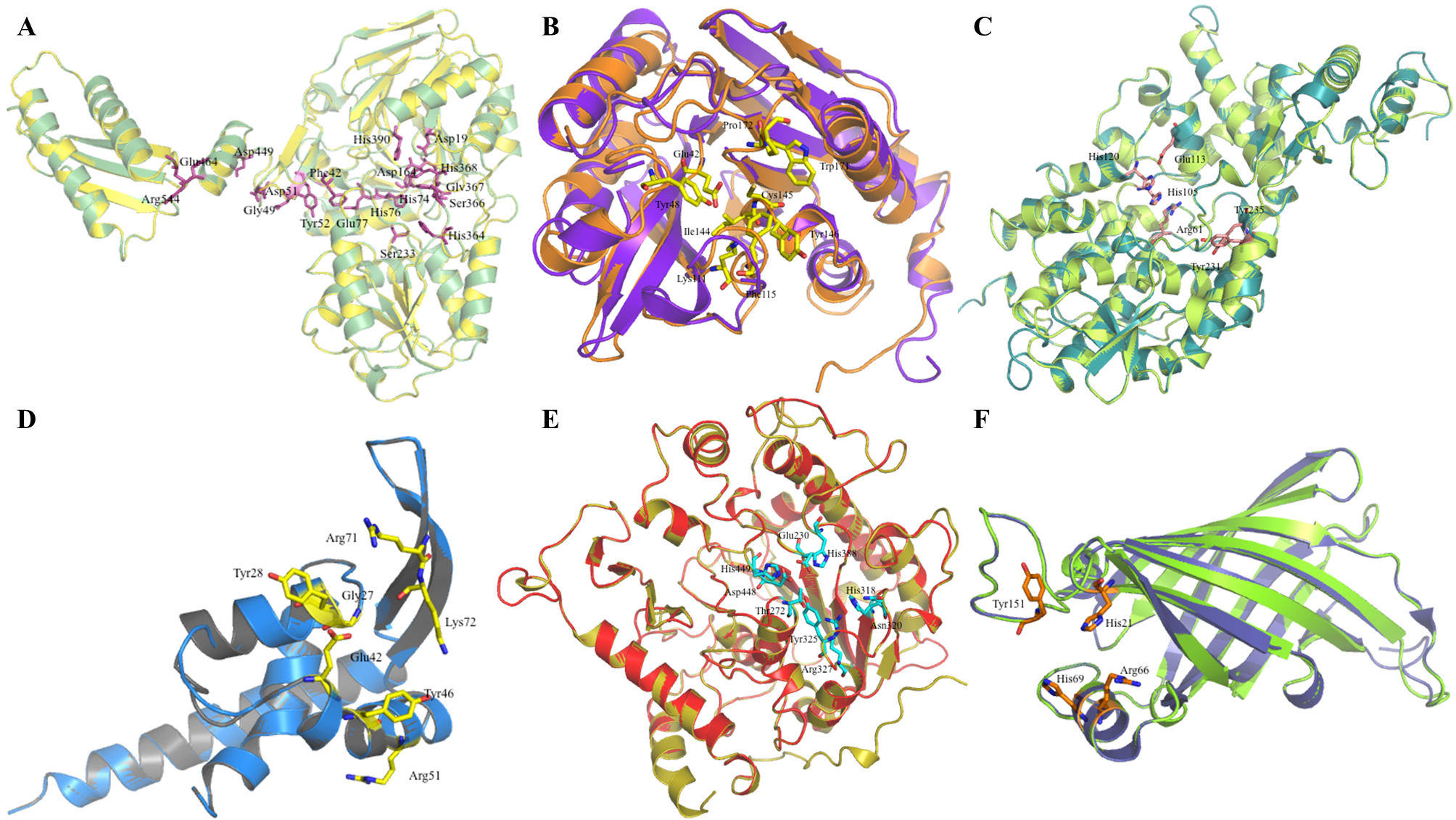
Figure 3. Structural superimposition of modeled proteins C1L2E5-C1KY50 with their respective template structures. Cartoon representations depicting the structural alignment of modeled proteins with their corresponding templates. (A) C1L2E5 (red) superimposed on template 1S3L (green). (B) C1KZM7 (raspberry) superimposed on template 3HGM (wheat). (C) C1KYZ6 (red) superimposed on template 3FTJ (green). (D) C1KYT3 (green) superimposed on template 1VI7 (red). (E) C1KY51 (pink) superimposed on template 1SU0 (blue). (F) C1KYL6 (blue) superimposed on template 3R4C (pink). (G) C1KY50 (red) superimposed on template 2ZU0 (cyan).
Download: Download full-size image
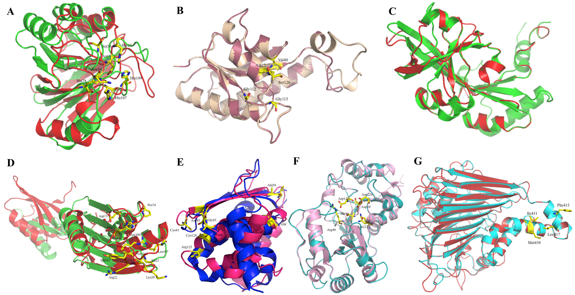
Table 3. Superimposed residues between template and modeled proteins C1L1U8-C1L165.
| Protein ID | Superimposed Residues (Template -> Model) |
|---|---|
| C1L1U8 | Asp78->Asp78, His79->His79, Asp164->Asp164, His390->His390, His74->His74, His76->His76, His142->His142, Asp195->Asp195, His368->His368, Gly49->Gly49, Asp51->Asp51, Asp443->Asp443, Glu464->Glu464, Asp449->Asp449, Arg544->Arg544, Ser366->Ser366, His364->His364, Gly367->Gly367, Ser233->Ser233, Glu77->Glu77, Phe42->Phe42, Tyr52->Tyr52 |
| C1KYM2 | Glu43->Glu42, Lys109->Lys111, Cys143->Cys145, Tyr144->Tyr146, Phe49->Tyr48, Phe113->Phe115, Trp175->Trp171, Val142->Ile144, Pro176->Pro172 |
| C1KYY1 | His129->His120, Glu122->Glu113, His66->His64, His110->His110, Asp111->Asp102, Asp183->Asp173, Lys14->Lys12, Asn36->Ala34, Gln41->Gln39, Arg326->Arg317, Lys330->Arg321, His114->His105, Tyr239->Tyr231, Arg63->Arg61, Leu49->Leu47, His119->His110, Tyr187->Tyr177, Tyr243->Tyr235, Tyr368->Tyr358 |
| C1L0M8 | Gly25->Gly27, Tyr26->Tyr28, Glu42->Glu42, Arg71->Arg71, Lys72->Lys72, Tyr46->Tyr46, Arg51->Arg51 |
| C1L0R8 | Thr297->Thr272, His412->His388, Glu253->Glu230, Trp350->Tyr325, His472->His449, Asp471->Asp448, His343->His318, Asn345->Asn320, Arg352->Arg327, Thr408->Ser384 |
| C1L165 | His18->His21, Arg62->Arg66, His65->His69, Trp146->Tyr151 |
Table 4. Superimposed residues between template and modeled proteins C1L2E5-C1KY50.
| Protein ID | Superimposed Residues (Template -> Model) |
|---|---|
| C1L2E5 | Asp8->Asp8, His10->His10, Asp36->Ser36, Asn59->Asn54, Asn60->Cys55, His97->His78, His120->His107, Thr121->Ser108, His122->His109 |
| C1KZM7 | Arg135->Tyr130, Ser131->Ser126, Val38->Val40, Pro8->Gly10, Gly117->Gly112, Gln119->Thr114, Ala133->Ser128, Val132->Val127, Gly120->Gly115, Gly123->Ala118, Asn122->Ser117 |
| C1KYZ6 | Leu469->Lys186, Tyr465->Trp182, Asn346->Ala79, Ile342->Thr75 |
| C1KYT3 | Ser23->Ser21, His54->His52, Glu77->Glu74, Arg104->Arg100, Lys22->Lys20, Arg24->Arg22, Phe25->Phe23, Asp75->Asp71, Gly76->Gly72, Pro78->Pro74, Ala82->Ala78, Tyr105->Tyr101, Tyr106->Phe102, Gly107->Gly103, Leu111->Leu107, Leu116->Leu112, Tyr120->Tyr116, Asp74->Asp70, Thr81->Thr77 |
| C1KY51 | Cys40->Cys41, Asp42->Asp43, Cys65->Cys66, Arg124->Arg125, Cys127->Cys128, Gly64->Gly65, Asp57->Ala58, Asp77->Gln78, Glu56->Val57 |
| C1KYL6 | Trp171->Ser187, Phe175->Asn191, Arg45->Arg46, Asp1->Asp12, Thr43->Thr44, Asp8->Asp10, Lys185->Lys201, Asn211->Asn227, Asp208->Asp22 |
| C1KY50 | Phe373->Phe415, Leu375->Leu417, Ile380->Leu422, Met338->Met430, Ile389->Ile431, Ala392->Gly434, Ala395->Glu437 |
4. Conclusions
We have conducted an extensive analysis of the hypothetical proteins of L. monocytogenes. Following genome analysis of pathogens, we observed numerous proteins whose functions remain uncharacterized. Comparative genomics indicates that these proteins are present across organisms but have not yet been functionally characterized to date. The first part of this study comprises proteins for which biochemical activity can be predicted with reasonable confidence using genomic sequence-based approaches. We listed the proposed functions of most HPs using sequence analysis tools. The second part includes structure determination and the development of a structure-function relationship. We have successfully determined the structure-function relationship for 13 HPs from Listeria monocytogenes. This structural genomics project enables us to better understanding of pathogenesis of this microorganism. We have finally achieved our goal of assigning structure-based biochemical functions to most hypothetical proteins from Listeria monocytogenes. However, further biochemical work is needed to assign functions with accurate precision.
5. Conflict of Interest:
There is no conflict of interest to declare.
6. Data Availability Statement:
The data supporting this study are provided in this article.
7. Funding:
None
8. Acknowledgements:
None
9. Supplementary materials:
None
References
Chin, K. H., Tsai, Y.-D., Chan, N. L., Huang, K. F., Wang, A. H. J., & Chou, S. H. (2007). The crystal structure of XC1258 from Xanthomonas campestris: A putative procaryotic Nit protein with an arsenic adduct in the active site. Proteins, 69(3), 665–671. https://doi.org/10.1002/PROT.21501
Fibriansah, G., Kovacs, A. T., Pool, T. J., Boonstra, M., Kuipers, O. P., & Thunnissen, A. M. W. H. (2012). Crystal structures of two transcriptional regulators from Bacillus cereus define the conserved structural features of a PadR subfamily. PLOS ONE, 7(11), Article e48015. https://doi.org/10.1371/JOURNAL.PONE.0048015
Galperin, M. Y., & Koonin, E. V. (2004). “Conserved hypothetical” proteins: Prioritization of targets for experimental study. Nucleic Acids Research, 32(18), 5452–5463. https://doi.org/10.1093/NAR/GKH885
Gebremedhin, E. Z., Hirpa, G., Borana, B. M., Sarba, E. J., Marami, L. M., Kelbesa, K. A., Tadese, N. D., & Ambecha, H. A. (2021). Listeria species occurrence and associated factors and antibiogram of Listeria monocytogenes in beef at abattoirs, butchers, and restaurants in ambo and holeta in ethiopia. Infection and Drug Resistance, 14, 1493–1504. https://doi.org/10.2147/IDR.S304871
Gury, J., Barthelmebs, L., Tran, N. P., Divies, C., & Cavin, J. F. (2004). Cloning, deletion, and characterization of PadR, the transcriptional repressor of the phenolic acid decarboxylase-encoding padA gene of Lactobacillus plantarum. Applied and Environmental Microbiology, 70(4), 2146–2153. https://doi.org/10.1128/AEM.70.4.2146-2153.2004
Hishigaki, H., Nakai, K., Ono, T., Tanigami, A., & Takagi, T. (2001). Assessment of prediction accuracy of protein function from protein–protein interaction data. Yeast, 18(6), 523–531. https://doi.org/10.1002/yea.706
Liu, J., Oganesyan, N., Shin, D. H., Jancarik, J., Yokota, H., Kim, R., & Kim, S. H. (2005). Structural characterization of an iron-sulfur cluster assembly protein IscU in a zinc-bound form. Proteins, 59(4), 875–881. https://doi.org/10.1002/PROT.20421
Lubec, G., Afjehi-Sadat, L., Yang, J. W., & John, J. P. P. (2005). Searching for hypothetical proteins: Theory and practice based upon original data and literature. Progress in Neurobiology, 77(1–2), 90–127. https://doi.org/10.1016/j.pneurobio.2005.10.001
Newman, J. A., Hewitt, L., Rodrigues, C., Solovyova, A., Harwood, C. R., & Lewis, R. J. (2011). Unusual, dual endo- and exonuclease activity in the degradosome explained by crystal structure analysis of RNase J1. Structure, 19(9), 1241–1251. https://doi.org/10.1016/j.str.2011.06.017
Nuesch-Inderbinen, M., Bloemberg, G. V., Muller, A., Stevens, M. J. A., Cernela, N., Kolloffel, B., & Stephan, R. (2021). Listeriosis caused by persistence of Listeria monocytogenes serotype 4b sequence type 6 in cheese production environment. Emerging Infectious Diseases, 27(1), 284. https://doi.org/10.3201/EID2701.203266
Sivashankari, S., & Shanmughavel, P. (2006). Functional annotation of hypothetical proteins - a review. Bioinformation, 1(8), 335–338. https://doi.org/10.6026/97320630001335
Vorontsov, I. I., Minasov, G., Kiryukhina, O., Brunzelle, J. S., Shuvalova, L., & Anderson, W. F. (2011). Characterization of the deoxynucleotide triphosphate triphosphohydrolase (dNTPase) activity of the EF1143 protein from Enterococcus faecalis and crystal structure of the activator-substrate complex. Journal of Biological Chemistry, 286(38), 33158–33166. https://doi.org/10.1074/jbc.M111.250456
Wada, K., Sumi, N., Nagai, R., Iwasaki, K., Sato, T., Suzuki, K., Hasegawa, Y., Kitaoka, S., Minami, Y., Outten, F. W., Takahashi, Y., & Fukuyama, K. (2009). Molecular dynamism of Fe-S cluster biosynthesis implicated by the structure of the SufC2-SufD2 complex. Journal of Molecular Biology, 387(1), 245–258. https://doi.org/10.1016/j.jmb.2009.01.054