


About Article
Investigation of inhibitory potential of resveratrol to the microtubule affinity regulating kinase 4: Implications in anticancer therapy
Volume 1, No. 1 · 2026
Highlights
- The study evaluates resveratrol as a natural inhibitor of microtubule affinity regulating kinase 4 (MARK4), a kinase implicated in cancer progression and neurodegeneration.
- Blind docking against the MARK4 crystal structure (PDB ID: 5ES1) suggested that resveratrol occupies the catalytic pocket and interacts through hydrogen bonds, hydrophobic contacts, and π–alkyl interactions.
- Fluorescence quenching experiments supported direct binding between resveratrol and MARK4, with an estimated binding constant of 7.0 × 10^4 M−1.
- MARK4 enzymatic activity decreased in a dose-dependent manner in the ATPase assay, and the reported IC50 for resveratrol was approximately 7 µM.
- Resveratrol also showed cytotoxicity against H1299 lung cancer cells, with an IC50 of approximately 114 µM in the cell proliferation assay.
Abstract
Resveratrol is presented as a bioactive natural compound with antioxidant, anticancer, and neuroprotective properties relevant to cancer progression and neurodegenerative disease.
The study examined resveratrol as an inhibitor of MARK4 using both in silico and in vitro approaches. Molecular docking was used to explore binding and structural compatibility of the ligand with MARK4.
Experimental follow-up included fluorescence binding studies and kinase inhibition analysis, which showed notable interaction and functional suppression of MARK4 activity.
The article reports an IC50 value of 7 µM for inhibition of MARK4 and a fluorescence-derived binding constant on the order of 10^4 M−1.
Overall, the findings support resveratrol as a promising MARK4-binding phytochemical scaffold with implications for anticancer therapy.
Keywords
Article Overview
This article examines the inhibitory potential of resveratrol against MARK4, a kinase linked to cytoskeletal regulation, tumor progression, and neurodegenerative disease.
The introduction connects dysregulated kinase signaling to cancer biology and argues that MARK4 is a relevant non-canonical kinase target because it influences cell-cycle progression, microtubule dynamics, polarity, metastasis-associated phenotypes, and pathological tau phosphorylation.
The authors position natural products as structurally versatile kinase modulators and describe resveratrol as a plausible MARK4-targeting phytochemical because of its known antioxidant, anti-inflammatory, neuroprotective, and anticancer activities.
1. About the Article
This is a research article published in Clinical & Molecular Biomedicine. It studies resveratrol as a potential inhibitor of MARK4 using docking, fluorescence binding, kinase inhibition, and cell proliferation assays.
The work combines computational and experimental approaches and frames MARK4 inhibition as relevant to both cancer and neurodegenerative disease.
- Journal: Clinical & Molecular Biomedicine
- Volume/Issue: Vol. 1, No. 1
- Header year: 2026
- Title: Investigation of inhibitory potential of resveratrol to the microtubule affinity regulating kinase 4: Implications in anticancer therapy
- Authors: Khuzin Dinislam; Minhal Abidi; Pratibha Prasad
- Affiliation 1: Bashkir State Medical University, Department of General Chemistry, Ufa, Republic of Bashkortostan, Russia
- Affiliation 2: Department of Biochemistry, Jawaharlal Nehru Medical College, Aligarh Muslim University, Aligarh, India
- Affiliation 3: Department of Basic Medical Sciences, Ajman University, United Arab Emirates
- Corresponding author: Pratibha Prasad
- Corresponding email: p.prasad@ajman.ac.ae
- Received: April 16th, 2024
- Accepted: April 1, 2026
- Published: May 21, 2024
- License: Creative Commons CC-BY 4.0
- Open-access status: Open Access
- Header page range on first page: pp. 1–25
- Printed article page range visible in PDF pages: 1–5
2. Introduction
The introduction describes protein kinases as central regulators of cellular signaling and highlights their relevance to malignant transformation, metastasis, and therapy resistance.
MARK kinases are introduced as AMPK-family members involved in microtubule stability, cytoskeletal reorganization, vesicle trafficking, polarity, and mitotic spindle organization. MARK4 is emphasized as a particularly important isoform because of its association with cancer, neurodegeneration, and metabolic imbalance.
The paper argues that selective inhibition of MARK4 could disrupt both structural and signaling components of tumor progression and reviews prior work suggesting that natural compounds such as rosmarinic acid, myricetin, and bacopaside II can modulate MARK4.
Resveratrol is then proposed as a suitable candidate because of its pleiotropic signaling effects and documented antioxidant, anti-inflammatory, neuroprotective, and anticancer activities.
- Target protein: microtubule affinity regulating kinase 4 (MARK4)
- Target class: kinase, MARK subfamily of AMPK-related kinases
- Therapeutic areas discussed: cancer, neurodegeneration, Alzheimer’s disease
- Natural-product rationale: phytochemical kinase modulation
- Lead compound studied: resveratrol (RV)
3. Materials and Methods
The methods combine molecular docking with protein expression and purification, ATPase-based enzyme inhibition, fluorescence binding studies, and H1299 cell proliferation analysis.
MARK4 docking used the human crystal structure from the PDB (5ES1). Protein preparation included removal of co-crystallized ligands and water molecules, hydrogen addition, and charge assignment before blind docking with InstaDock.
Experimental work included recombinant MARK4 expression in E. coli M15 cells using a pQE30 vector system, enzyme inhibition analysis with BIOMOL green reagent, fluorescence quenching measurements on a Jasco spectrofluorometer, and MTT-based cell viability analysis in H1299 lung cancer cells.
- Protein structure source: RCSB PDB ID 5ES1
- Docking software: InstaDock
- Visualization software: Discovery Studio Visualizer; PyMOL
- Docking mode: blind docking
- Docking exhaustiveness: 8
- Expression host: E. coli M15
- Expression vector: pQE30
- Final MARK4 concentration in inhibition assay: 5 µM
- Final ATP concentration in inhibition assay: 200 µM
- Fluorescence assay temperature: 25 °C
- Fixed MARK4 concentration in fluorescence assay: 4 µM
- Fluorescence excitation wavelength: 280 nm
- Fluorescence emission range: 300–400 nm
- Cell line: H1299 human non-small cell lung carcinoma
2.1. Materials
The article lists LB broth, IPTG, kanamycin, ampicillin, CAPS buffer, NaCl, glycerol, and resveratrol stock preparation conditions.
Resveratrol was prepared as stock solutions in an appropriate solvent such as DMSO and stored according to the manufacturer’s guidance.
2.2. Molecular Docking
Human MARK4 structure 5ES1 was prepared for docking by removing bound ligands and waters and assigning hydrogens and Kollman charges.
Blind docking was performed with InstaDock and analyzed using Discovery Studio Visualizer and PyMOL.
2.3. Protein expression and purification
MARK4 was expressed in E. coli M15 cells carrying pREP4/lacI support for pQE-based regulation.
The recombinant pQE30 system included an N-terminal polyhistidine tag to facilitate purification.
2.4. Enzyme inhibition assay
The assay measured phosphate release from ATP in the presence of MARK4 and increasing ligand concentrations, using BIOMOL green reagent for detection.
Absorbance was measured at 620 nm after color development.
2.5. Fluorescence binding studies
Intrinsic fluorescence quenching of MARK4 was monitored upon titration with RV to assess binding behavior and estimate binding parameters through modified Stern–Volmer analysis.
The study reports the standard Stern–Volmer and modified Stern–Volmer equations in the methods.
2.6. Cell proliferation assay
H1299 NSCLC cells were seeded in 96-well plates, treated with increasing RV concentrations, and evaluated using an MTT-based assay.
Absorbance was recorded at 570 nm after incubation with MTT.
4. Results and Discussion
Docking suggested that RV occupies the MARK4 catalytic pocket and interacts with residues Ile62 and Glu133 through conventional hydrogen bonds, along with multiple hydrophobic, van der Waals, and π–alkyl contacts.
Fluorescence binding studies showed progressive quenching with increasing RV concentration and were interpreted as evidence of direct binding with an estimated association constant of 7.0 × 10^4 M−1.
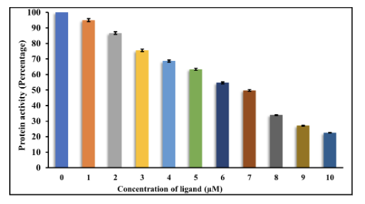
In the kinase assay, MARK4 activity decreased as RV concentration increased, and the reported IC50 was approximately 7 µM.
In H1299 cells, RV reduced cell proliferation over 48 hours and the reported IC50 was approximately 114 µM.
- Docking interaction site: catalytic pocket of MARK4
- Fluorescence binding constant: 7.0 × 10^4 M−1
- MARK4 inhibition IC50: ≈ 7 µM
- H1299 cell proliferation IC50: ≈ 114 µM
3.1. Molecular docking analysis
RV was reported to form hydrogen bonds with Ile62 and Glu133 inside the MARK4 binding cavity.
Additional interactions involved Met132, Val116, Tyr134, Gly138, Glu139, Asp142, Ala135, Leu185, and π–alkyl contacts with Val70 and Ala83.
- Hydrogen-bond residues: Ile62; Glu133
- π–alkyl residues: Val70; Ala83
- Other pocket residues mentioned: Met132, Val116, Tyr134, Gly138, Glu139, Asp142, Ala135, Leu185
3.2. Fluorescence-based binding studies
The fluorescence emission intensity of MARK4 decreased with increasing RV concentration, suggesting perturbation of the tryptophan microenvironment after ligand binding.
Modified Stern–Volmer analysis produced a reported binding constant of 7.0 × 10^4 M−1.
- Observed trend: concentration-dependent quenching
- Reported binding constant: 7.0 × 10^4 M−1
3.3. Enzyme inhibition assay
The ATPase activity assay showed dose-dependent reduction in MARK4 activity upon RV treatment.
The paper reports an IC50 of about 7 µM for RV against MARK4.
- Assay readout: ATPase/phosphate-release assay
- Reported IC50: ≈ 7 µM
3.4. Cell proliferation assay
RV exhibited cytotoxicity against H1299 lung cancer cells relative to the DMSO vehicle control.
The cell survival data are summarized as an approximate IC50 of 114 µM after 48 hours.
- Cell line tested: H1299
- Reported IC50 against H1299: ≈ 114 µM
5. Conclusion
The article concludes that resveratrol is a promising phytochemical scaffold for MARK4 targeting because it shows structural compatibility in docking, measurable direct binding in fluorescence experiments, functional kinase inhibition, and cytotoxic effects in H1299 cells.
The authors further argue that MARK4–resveratrol interaction provides a useful starting point for rational drug design aimed at multi-pathway therapeutic intervention in cancer and related diseases.
The conclusion also notes translational challenges for natural compounds, including bioavailability, metabolism, and potency, and points to medicinal chemistry, structural biology, and nanotechnology-based delivery as future directions.
- Proposed lead scaffold: resveratrol
- Target: MARK4
- Application focus: cancer and related diseases
- Future work noted: structural dynamics, pharmacokinetic optimization, translational development
6. Statements and Declarations
The closing section includes statements for competing interests and data availability.
- Declaration of Competing Interest: The authors report no conflicts of interest.
- Data availability statement: All data generated or analyzed during this study are included in this article.
Figures
The PDF includes four named figures covering docking views, fluorescence binding, ATPase inhibition, and cell proliferation analysis.
Figure 1


Figure 1. Molecular docking of RV with MARK4; 3D and 2D interaction views.
Download figure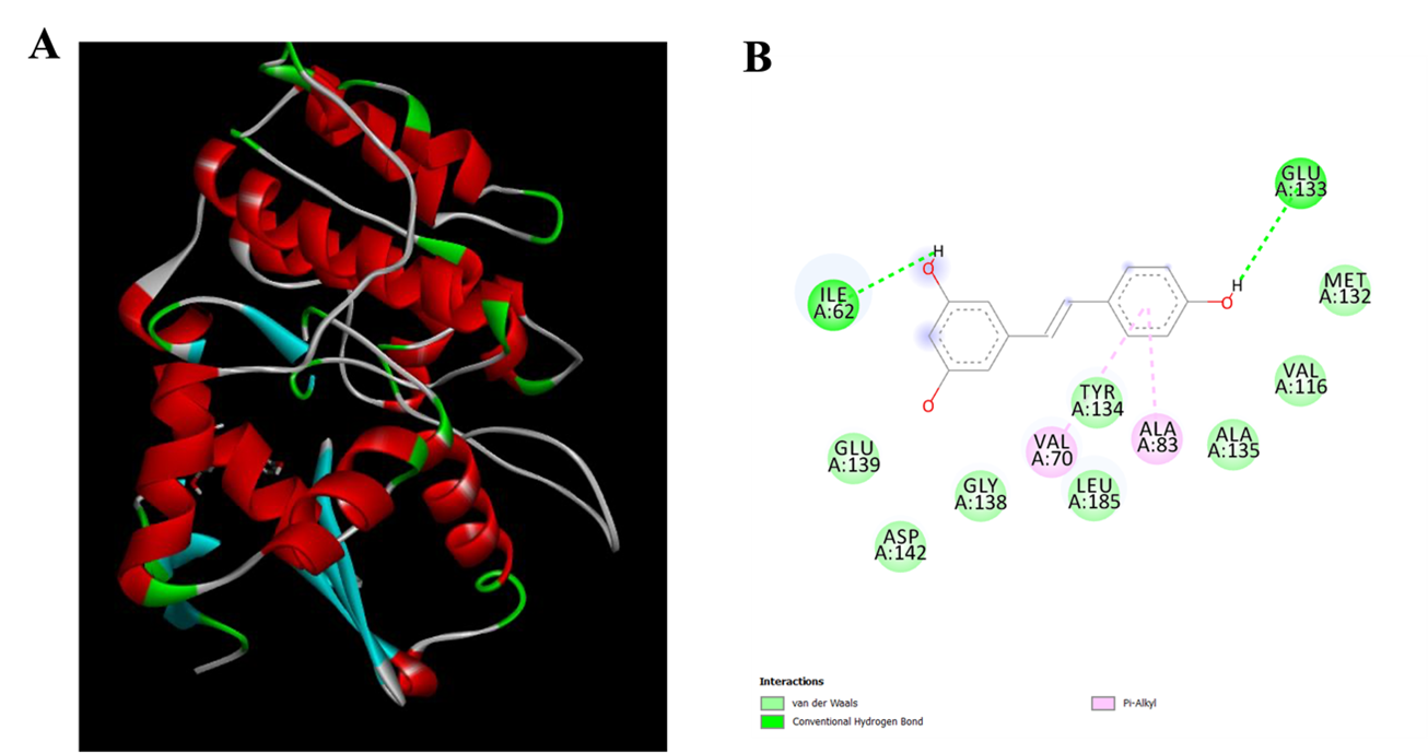

Figure 2. Fluorescence emission spectra of MARK4 with increasing RV concentration and modified Stern-Volmer plot.
Download figure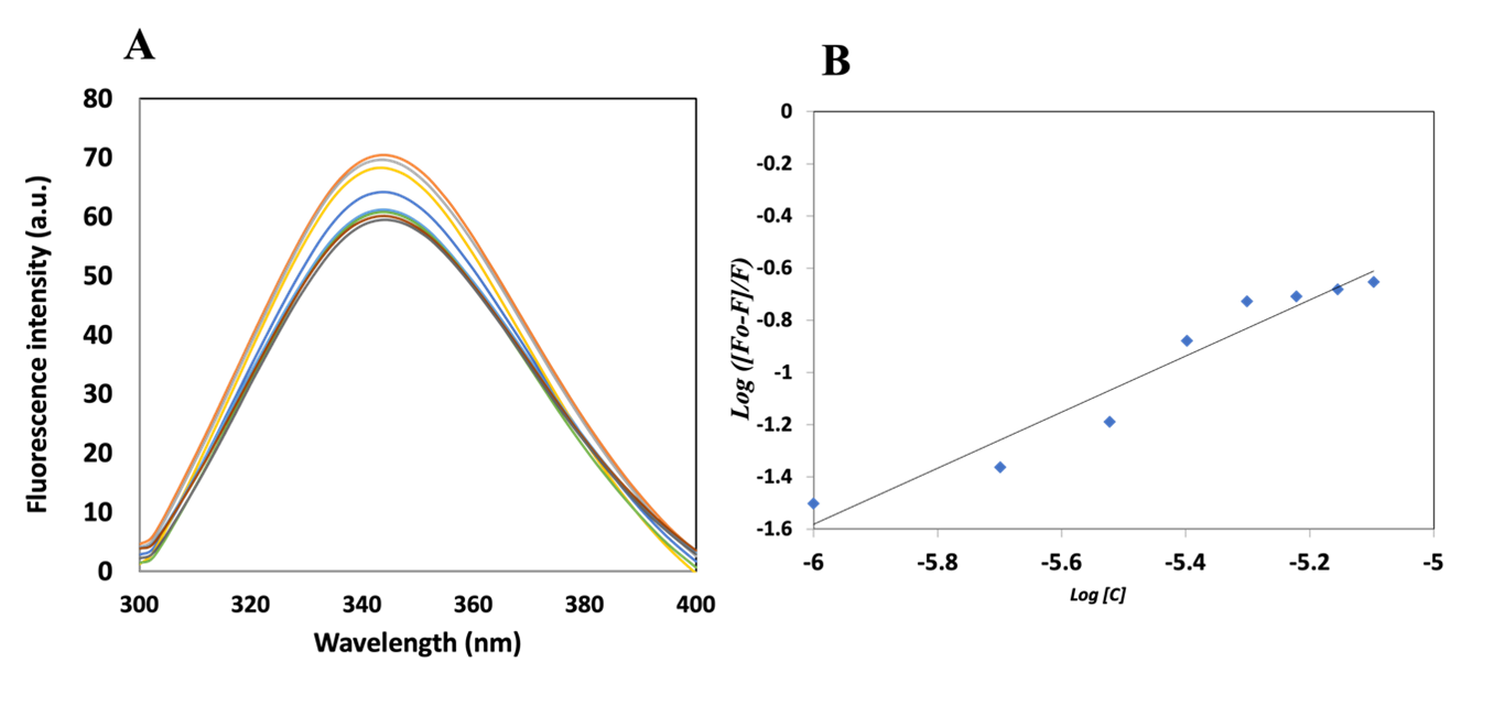

Figure 3. ATPase activity assay of MARK4 with varying RV concentration (0–10 µM).
Download figure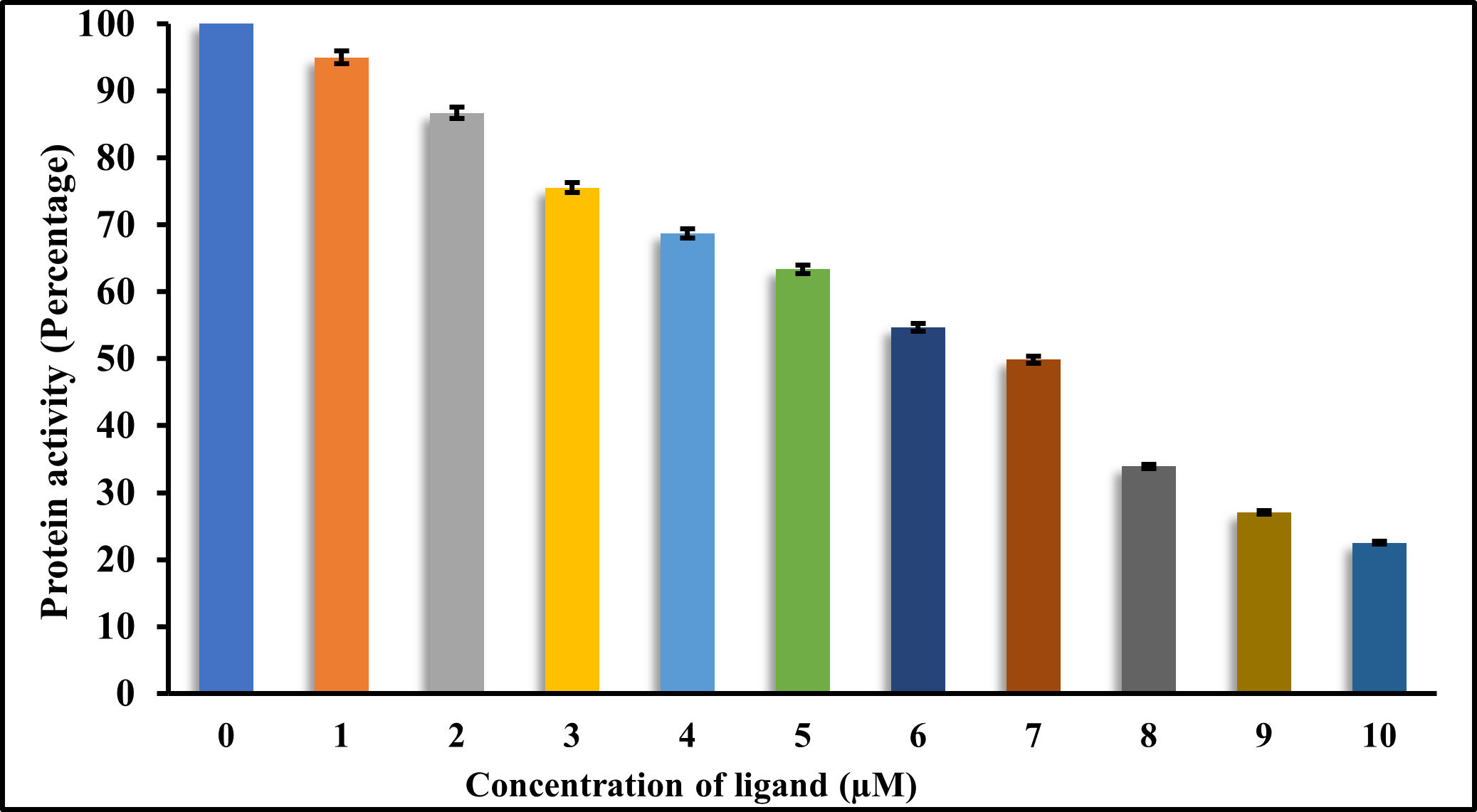

Figure 4. Cell proliferation assay of RV against H1299 cell line.
Download figure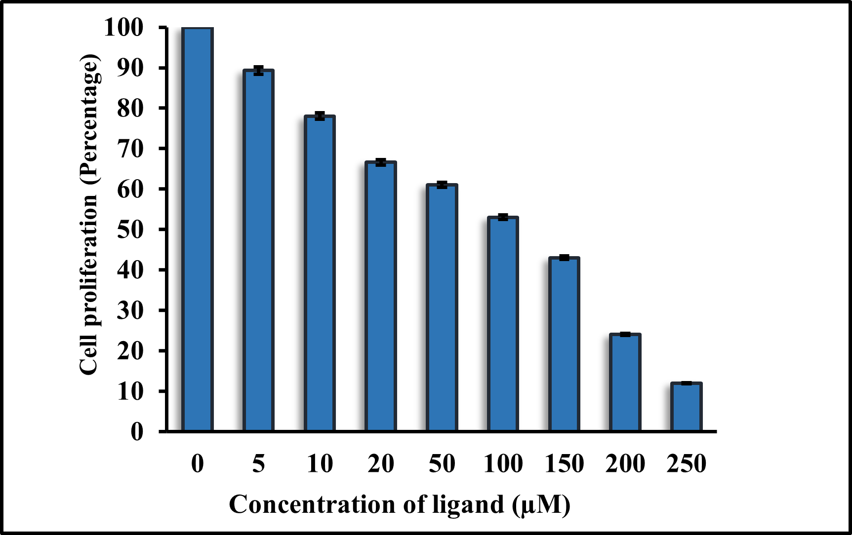
References
- 1
Alrouji, M., et al.
International Journal of Biological Macromolecules article cited in the paper.
International Journal of Biological Macromolecules · 2023 - 2
Anwar, S., et al.
Journal of Molecular Liquids article cited in the paper.
Journal of Molecular Liquids · 2022 - 3
Atiya, A., et al.
Journal of Biomolecular Structure and Dynamics article cited in the paper.
Journal of Biomolecular Structure and Dynamics · 2023 - 4
BIOVIA, Dassault Systèmes.
Discovery Studio Visualizer.
Software reference · 2017 - 5
Dahiya, R., et al.
RSC Advances article cited in the paper.
RSC Advances · 2019 - 6
DeLano, W. L.
CCP4 Newsletter on Protein Crystallography article cited in the paper.
CCP4 Newsletter on Protein Crystallography · 2002 - 7
Eftink, M. R.
Methods in Enzymology article cited in the paper.
Methods in Enzymology · 1997 - 8
Fan, J., Fu, A., & Zhang, L.
Quantitative Biology article cited in the paper.
Quantitative Biology · 2019 - 9
Lakowicz, J. R.
Principles of Fluorescence Spectroscopy.
Book · 2006 - 10
Lakowicz, J. R., & Weber, G.
Biochemistry article cited in the paper.
Biochemistry · 1973 - 11
Mohammad, T., Mathur, Y., & Hassan, M. I.
Briefings in Bioinformatics article cited in the paper.
Briefings in Bioinformatics · 2021 - 12
Shamsi, A., et al.
Biomolecules article cited in the paper.
Biomolecules · 2020 - 13
Taraska, J. W., & Zagotta, W. N.
Neuron article cited in the paper.
Neuron · 2010 - 14
Valeur, B., & Berberan-Santos, M. N.
Molecular Fluorescence: Principles and Applications.
Book · 2013 - 15
Vivian, J. T., & Callis, P. R.
Biophysical Journal article cited in the paper.
Biophysical Journal · 2001